


2018 kavli prize in Neuroscience
2018 Kavli
Prize in
Neuroscience
The Norwegian Academy of Science and Letters has decided to award the 2018 Kavli Prize in Neuroscience to Robert Fettiplace, A. James Hudspeth and Christine Petit.


"For their pioneering work on the molecular and neural mechanisms of hearing.”
Committee Members
- Ole Petter Ottersen (Chair), Centre for Molecular Biology and Neuroscience, University of Oslo, Norway
- Catherine Dulac, Harvard University, USA
- Susan McConnell, Stanford University, USA
- Alexander Borst, Max Planck Institute of Neurobiology, Germany
- Antoine Triller, Institut de Biologie de l'École Normale Supérieure, France
Citation from the committee
Hearing is an important sense that contributes to human communication. The three Kavli Prize laureates used complementary approaches to unravel the mechanisms by which nerve cells transform sounds into electrical signals. This process is performed in the inner ear by sensory receptors called hair cells. The unique cellular, molecular and biophysical properties of these cells enable them to detect small air vibrations across a wide range of frequencies. The electrical signals generated by hair cells are then transmitted into the brain, allowing them to be interpreted as language, music, or noise.
A. James Hudspeth has provided the major framework for our understanding of the process that transduces sound into neural signals. Extending from each hair cell is a bundle of fine processes that act as sensors. Hudspeth used ingenious methods to reveal how sound-induced vibrations, which set the hair bundle in motion, evoke an electrical response in the hair cells through a direct mechanical connection between the hair bundle and ion channels. He also revealed how sound signals, which can be extremely small, are amplified within the inner ear.
Robert Fettiplace has made fundamental contributions to our understanding of sound transduction and demonstrated that each hair cell in the cochlea of the inner ear is sensitive to a specific range of sound frequencies. His experiments revealed that hair cells are organized along the cochlea in a pattern that reflects their frequency selectivity. Using sensitive physiological measurements and theoretical modeling, he discovered that this selectivity reflects an intrinsic electrical property of the cell, set by the density and kinetics of its ion channels that induce a resonance at a particular frequency.
Christine Petit has explored the genetics of hereditary deafness in humans and identified more than twenty genes that are required for hearing and inner ear development. She elucidated the mechanisms through which these mutations cause hearing deficits, thus illuminating the unique biology of hair cells and informing deafness diagnosis and counseling. Several of the genes she identified form major components of the hair cell mechanotransduction machinery. Collectively the breakthroughs made by this year’s Kavli Prize laureates have unveiled the molecular and cellular mechanisms that underlie hearing and deafness.
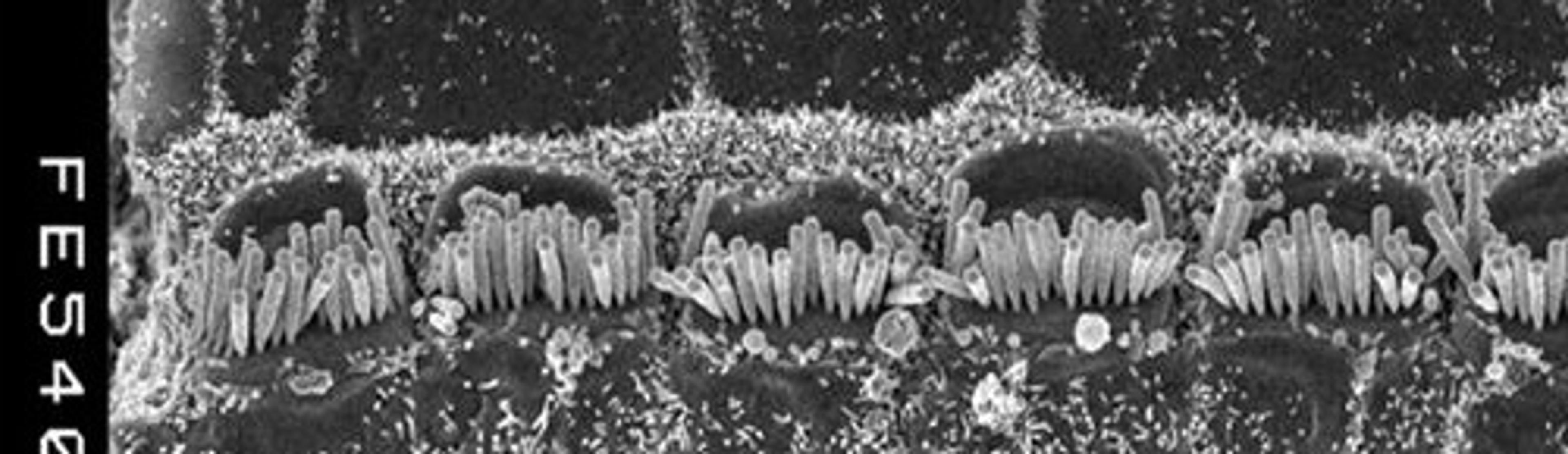
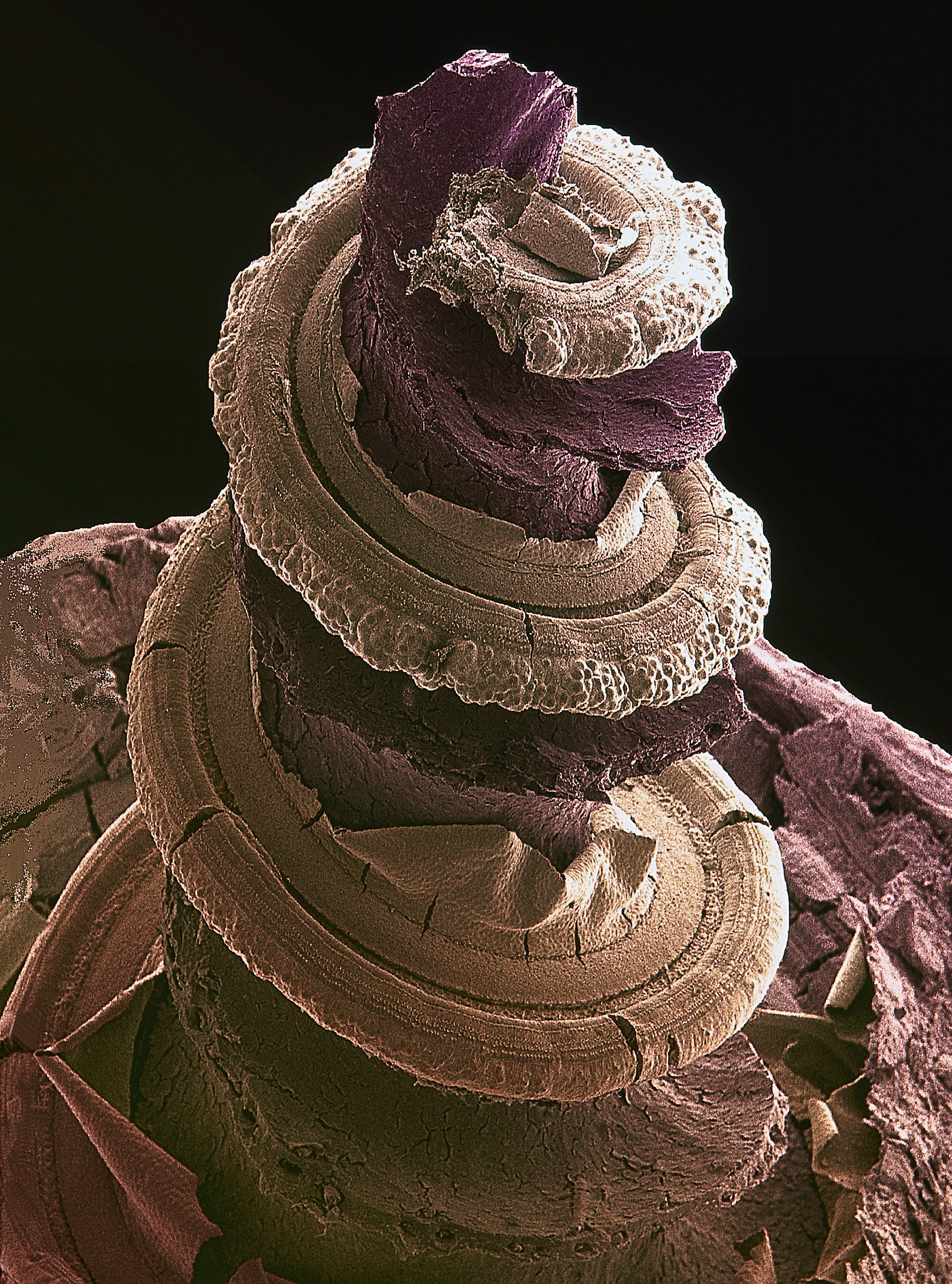
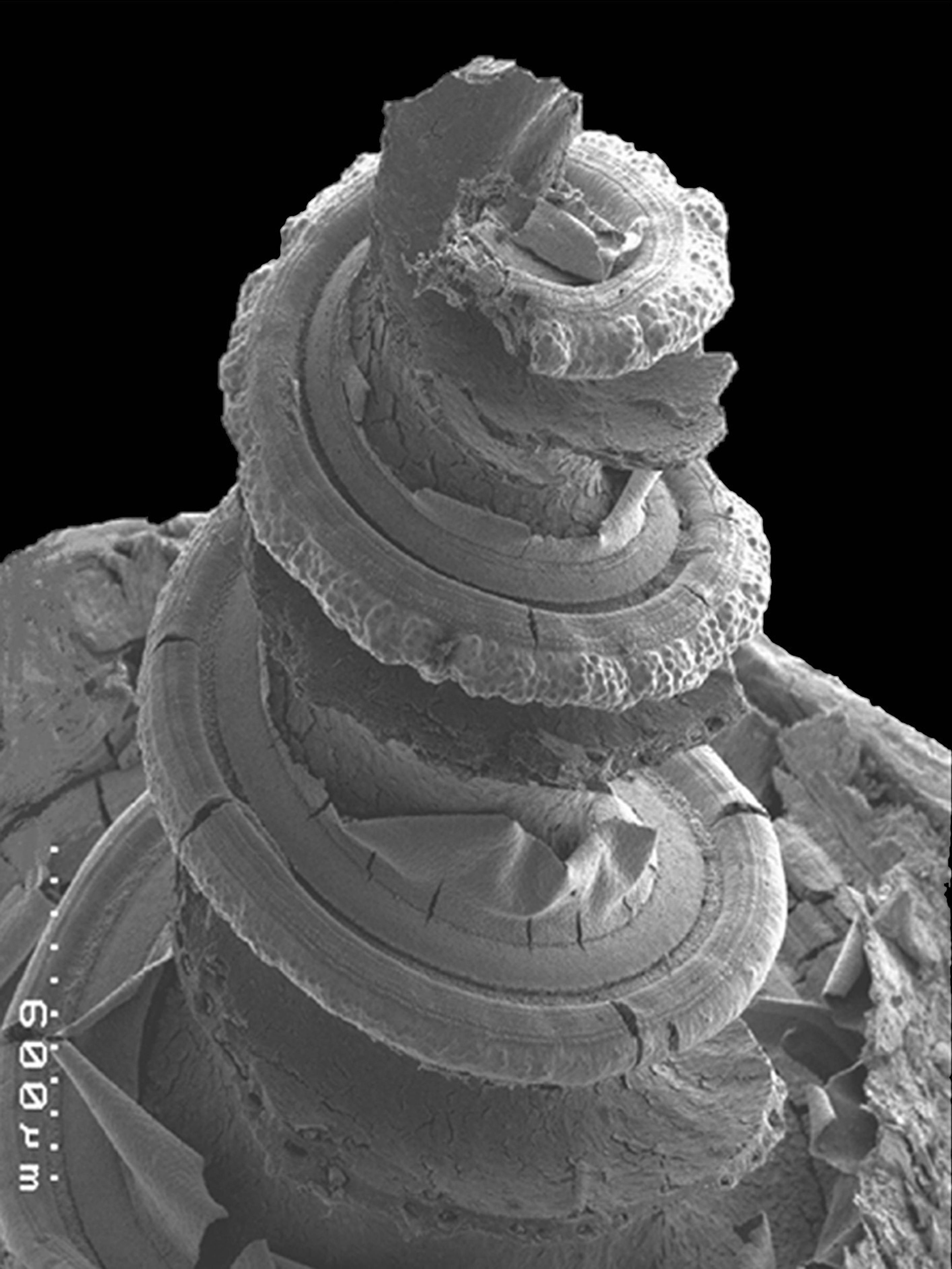
Along the spiral structure is the Organ of corti (right) that contains rows of sensory hair cells which respond to different frequencies of sound. The cells send impulsesalong the auditory nerve to the brain. (Photo credit: Dr. David Furness/Wellcome collection)
Tuning in to the mechanism of hearing
Listening to music, a person talking, or an animal rustling through leaves, all depend on our sensitive hearing. The human ear is as sensitive for sound as eyes are for a photon of light. But how do we distinguish sounds, whether faint or loud, distant or near?
By Julie Clayton
The 2018 Kavli Prize for Neuroscience has gone to scientists who have shed light on the fundamental processes of hearing, and helped to explain deafness.
Until a few decades ago, little was known about hearing beyond basic anatomy and physiology. Sound waves enter the ear canal of the outer ear and cause the eardrum to vibrate; the vibrations travel through the bones of the middle ear to the cochlea, in the inner ear. This tiny organ, shaped like a snail’s shell, is just a few millimeter wide, and 32 mm long in humans. Sound pressure waves travel through its fluid-filled chambers, and trigger electrical impulses to be sent to the brain via the auditory nerve. Scientists suspected that cells known as hair cells might be the key sensors of sound signals.

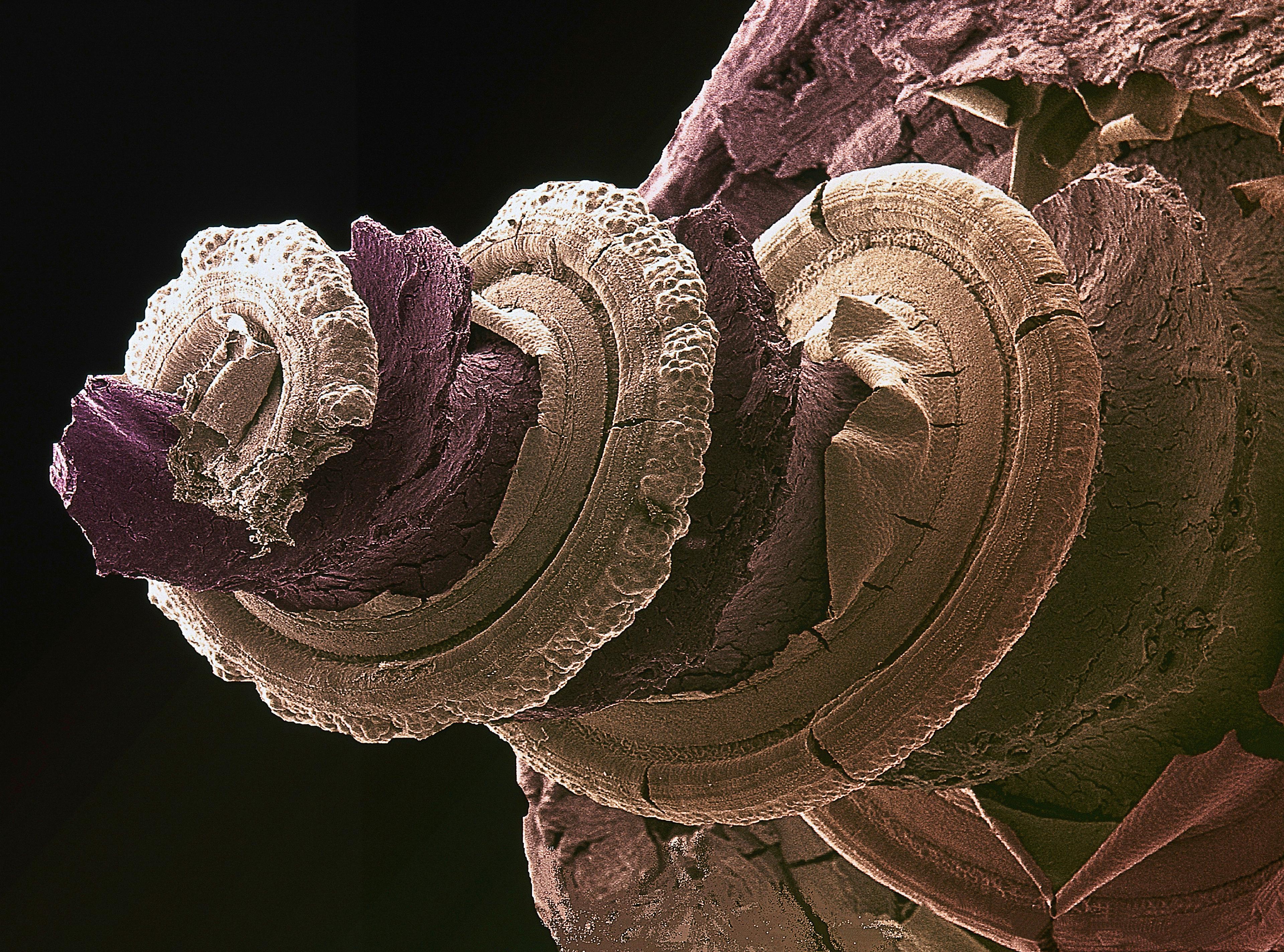
Colour-enhanced scanning electron micrograph of the inside of a guinea pig inner ear showing the hearing organ, or cochlea. Running along the spiral structure are rows of sensory cells which respond to different frequencies of sound. The whole organ is just a few millimeters long. (Photo credit: Dr. David Furness/Wellcome collection
There are only around 16,000 hair cells in the cochlea, lining the basilar membrane, which separates the fluid-filled ducts of the cochlea. Microscopy shows that over a lifetime, these can become damaged and lost following exposure to drugs such as streptomycin, infections such as rubella or toxoplasmosis, or loud sounds, and do not regenerate, which could account for hearing loss.
The 2018 Kavli Neuroscience prizewinners, James Hudspeth, Robert Fettiplace and Christine Petit, have independently investigated the role of hair cells in hearing. Hair cells are named for their tufts of hair-like projections visible on electron microscopy. These tufts, called hair bundles, consist of 20 to 300 individual projections called stereocilia, arranged in neat rows of different height, and are embedded in a jelly-like overlay called the tectorial membrane.
Starting in the late 1970s, Hudspeth and Fettiplace came from biophysics backgrounds, and were curious about whether movement of the hair bundles, due to vibrations of the cochlear membranes, led to electrical signaling. In order to access the cochlea – which in mammals is embedded inside the temporal bone of the skull – they both chose initially to work on cochlea from exotic animals such as bull frogs (Hudspeth) and turtles (Fettiplace). These are fairly large and easy to maintain in the laboratory because, coming from cold-blooded creatures they can withstand temperature fluctuations. They then had to delicately peel off the tectorial membrane without tearing the hair bundle structures.
Hudspeth fashioning a very fine glass fibre, with a diameter of 0.5 to 0.8 micrometers, with which to push gently against the tips of individual hair bundles. He used a piezoelectric actuator for precise control of the glass fibre, and equally fine microelectrodes to measure changes in electrical potential of individual hair cells. He was the first to show that mechanical displacement of a hair cell bundle in the direction of the tallest row of stereocilia, triggered a change in electrical charge (depolarization) of the cell membrane. The amount of displacement needed was so miniscule, that Hudspeth likened it to being the equivalent of a thumb-wide movement at the tip of the Eiffel Tower.
Hudspeth found that the electrical response required the flow of potassium and calcium ions through the membrane at tips of the stereocilia, and proposed that the movement of stereocilia mechanically pulls open membrane channels to allow positively charged ions to flow in.
He drew inspiration from electron microscopy images of short rod-like structures connecting neighbouring stereocilia Tip-links, he suggested, were stretched by the movement of stereocilia and exerted a force that opened the ion channels. When moved in the opposite direction, the tip-links slackened and the ion channels closed. This would be consistent with the idea that sound waves pulsating through the fluid ducts of the cochlea, cause the basilar and tectorial membranes to vibrate and move across one another, creating a shearing force on hair bundles.
For the next three decades, Hudspeth developed his system and revealed more of the biophysical and biochemical properties of hair cells and their putative ion channels. He revealed a phenomenon that was like a public address system being turned up too far: rather than being passive receivers of sound, hair bundles actively twitch, which confers a 100- to 1000-fold greater sensitivity to sound vibrations. In other words, have a built-in amplifier system. It is this amplification that is lost first with aging.

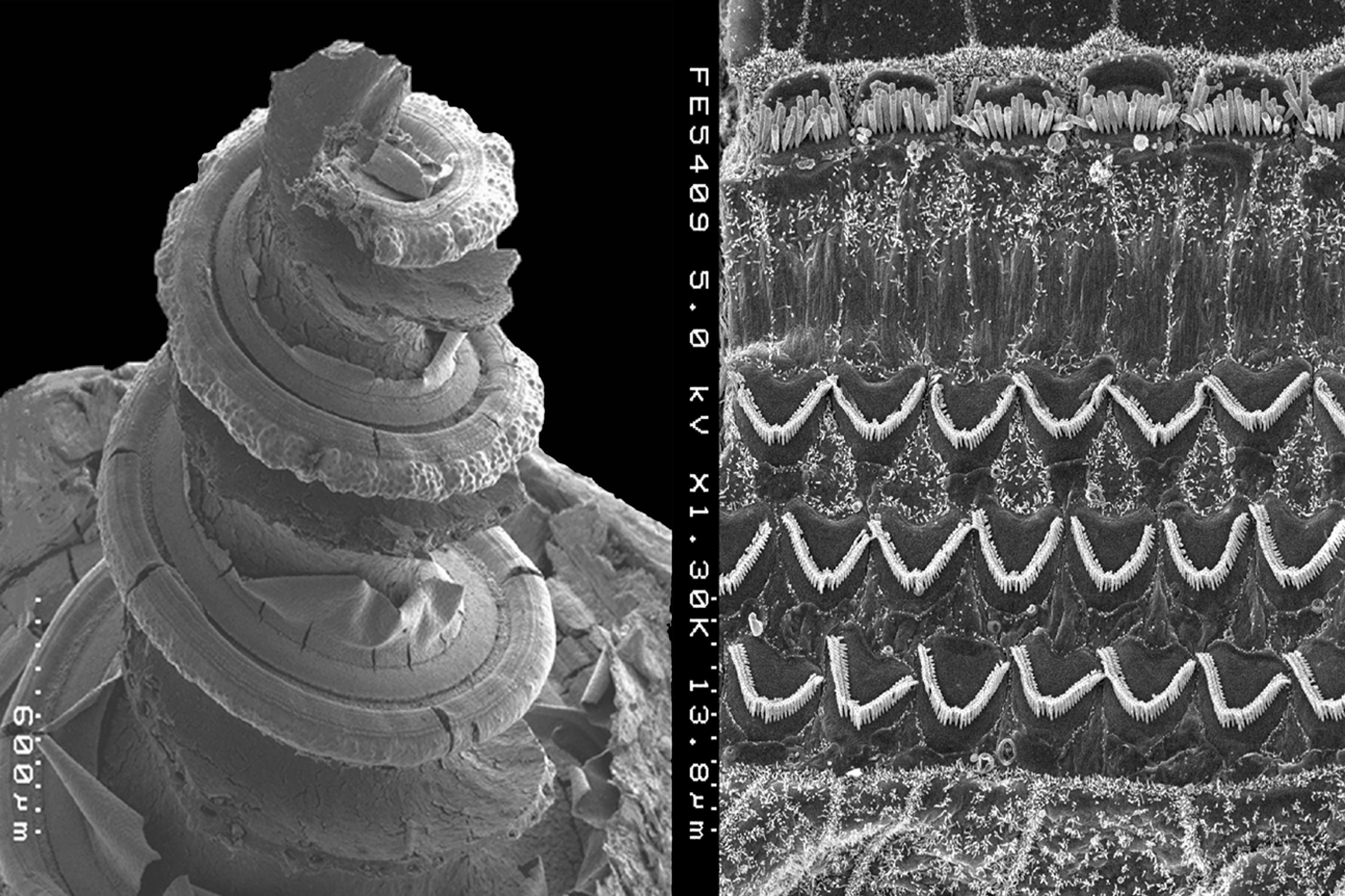
Cochlea of the inner ear. A scanning electron micrograph of the inside of a guinea pig inner ear showing the hearing organ, or cochlea (left). Along the spiral structure is the Organ of corti (right) that contains rows of sensory hair cells which respond to different frequencies of sound. The cells send impulses along the auditory nerve to the brain. (Photo credit: Dr. David Furness/Wellcome collection)
Robert Fettiplace investigated the turtle cochlea, and in 1980, reported that the sensitivity to different sound frequencies could be mapped along the basilar membrane – rather like the keys of a piano. By recording electrical changes in inner hair cells, he revealed a spectrum of sound sensitivity, called a tonotopic map, in which individual hair cells were tuned to resonate with just a narrow range of sound frequencies, according to their position along the membrane. Hair cells at the narrower, stiffer base of the cochlea, responded to high frequencies, while those at the wider and more floppy apex of the membrane responded to low frequencies. Fettiplace predicted – and over the next two decades showed experimentally - that the fine tuning of each hair cell is due to the numbers and speed of opening and closing of different ion channels, which influences the hair cell’s electrical and mechanical properties. Hair cells at the two ends of the cochlea differ in their composition of ion channels and the height of their hair bundles. The identity of the ion channels, and how they open and close, remain a mystery.
Mechanotransduction in hair cells of the inner ear. (A) Scanning electron micrograph of hair bundle (bullfrog sacculus; David P. Corey's Lab.). This top view shows the stereocilia arranged in order of increasing height. (B) Model for mechanotransduction. Deflection of a hair cell's bundle causes the stereocilia to bend and the tip links between them to tighten. (C) Ion channels attached to intracellular elastic elements (ankyrin repeats) open in response to tension on the rather inextensible tip link.
Fettiplace later found that in mammals, the amplification of sound sensitivity is due to the movements of outer hair cells, which enhance vibrations locally in the basilar membrane.
By the early 1990s, Hudspeth, Fettiplace and others had made major strides in understanding the biophysics and physiology of the auditory system, but little was known about the molecular mechanisms. This was especially difficult to study because the cochlea has so few hair cells specific to each sound frequency.
This is where Christine Petit’s research compliments that of Hudspeth and Fettiplace. Trained in medicine in France, she was interested in the genetic basis for inherited forms of deafness. Petit worked extensively to build collaborations with doctors in Syria, Lebanon, Algeria, Tunisia, Morocco and Jordan, where profound deafness appeared to be particularly prevalent in some large families. One form is Usher syndrome, featuring progressive blindness and hearing defects, and which Petit found involved several different genes. There are over a hundred such inherited syndromes, and many different genetic mutations involved.
Through genetics, molecular biology and biochemical analysis, Petit has identified over 20 distinct genes whose absence or mutation disrupts hearing, mainly by affecting the development and function of hair cells. Some encode proteins of the tip-link complex, the ankle-links at the base of the hair bundle, the top-connectors at its apex and machinery involved in the release of the neurotransmitter glutamate, and other hair cell components. There are hints too that some of the genes may also be involved in the wiring of the auditory cortex, where the brain decodes the information about sound.
Petit’s work pinpointed the proteins at the heart of the molecular machinery proposed by Hudspeth and Fettiplace, and has also provided an explanation of some of the hundreds of different forms of human hearing loss. The work of the 2018 prizewinners may even have a practical application in future through gene therapy, or regeneration of hair cells in the inner ear to replace those that become damaged over time.
Medical applications aside, the 2018 Kavli Prize in Neuroscience honours curiosity-driven basic research that advances our understanding of hearing. According to Christine Dulac, a member of the Kavli Prize Neuroscience Committee, the prizewinners demonstrate, “without any shadow of a doubt, that there is this inter-dependence between this basic research and clinical research. Those different realms of science are inextricably intertwined.”