


The Fruit of Real Teamwork
As told by Christine Petit
I was born in Laignes, a small village in northern Burgundy close to the sources of the Seine. This village, home to my father’s family, is located in a highly forested region. Its stone houses call to mind a golden age of forges and the industrial and domestic complex created by the great 18th century naturalist, the Comte de Buffon, embodies the spirit of the Age of Enlightenment. My mother came from southern Burgundy, with its sun-drenched hillsides and very famous wines. Her family had been established as vintners in Chassagne-Montrachet for generations. I was greatly influenced by my father, a brilliant physical engineer and pianist, and his passion for scientific discoveries. I was also marked by the frustration of my paternal grandmother, who, although an excellent pupil, was denied the chance to become a teacher, in an epoch in which women’s destinies were predetermined.

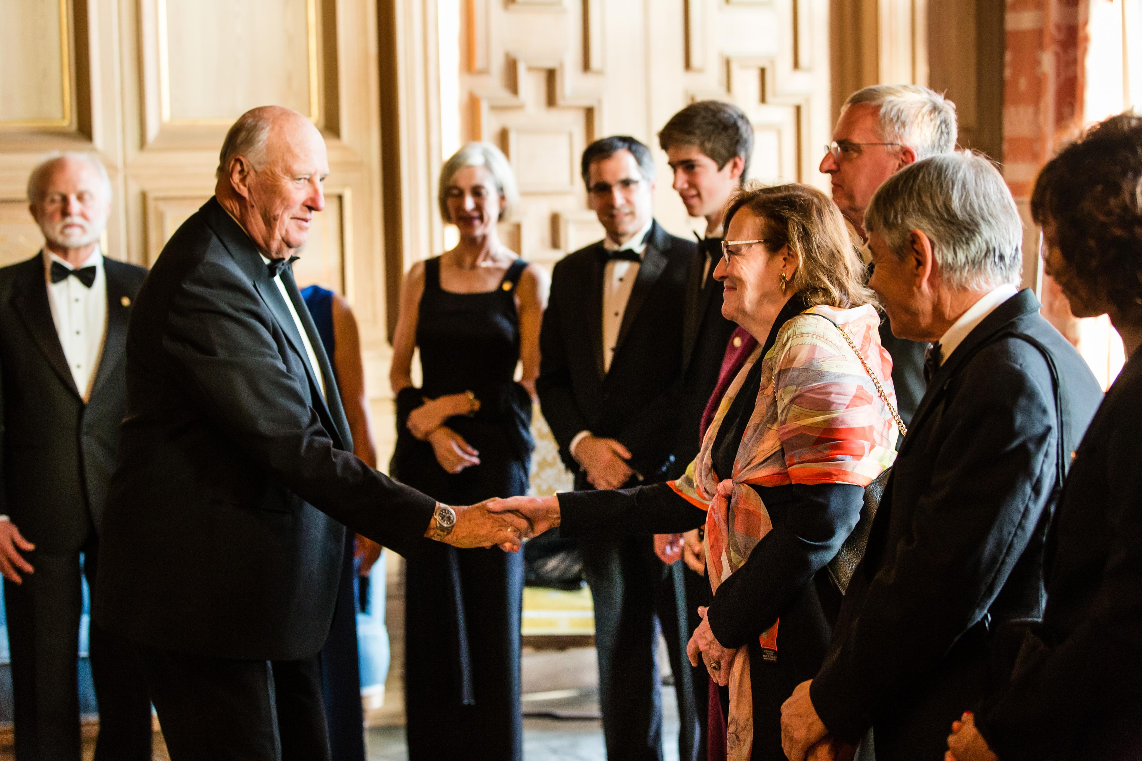
2018 Kavli Prize laureate Christine Petit meets His Royal Highness King Harald during the Kavli Prize week in Oslo (Photo credit: Thomas Eckhoff).
My interest in science began early. I trained as a physician at Paris VI University and Pitié-Salpétrière Hospital, a historic cradle of neurology. I soon realized that I would need additional basic science training to reach the depth of understanding I sought in biological science. I attended additional courses in parallel and graduated with a Masters in genetics and biochemistry from Paris XI University of Sciences at Orsay, in 1973. I was lucky enough to be taught by Georges Rizet, a pioneer of genetic training in France, who revealed to me the rational power and heuristic value of genetics. I took microbiology and virology courses at Institut Pasteur in Paris, in 1974, at the end of which I was offered a position at the Institute.
I initially worked towards my doctoral degree in science with Gunnar Lindahl in the laboratory of the Nobel laureate François Jacob, where I studied bacterial immunity to bacteriophages lambda and P2. I then moved to animmunology laboratory, where I obtained my doctorate in 1982. After a short stint at the Institute of Immunology in Basel, Switzerland, I moved to the CNRS center at Gif-sur-Yvette, where I focused on identifying genes regulating cell differentiation, through a genetic approach involving microcell hybrids. I returned to Institut Pasteur as a staff scientist in 1985.
A dream come true
At the time, new opportunities were emerging for deciphering human gene function and dysfunction by identifying disease-causing genes. This possibility, which had long been a dream for me, was finally becoming a reality, thanks to new methods for analyzing the human genome. I first addressed the molecular mechanisms underlying sex inversions in humans with my colleague Jacqueline Levilliers and Jean Weissenbach. We found that most cases of XX maleness and some cases of XY femaleness result from abnormal terminal exchanges between the short arms of the X and Y chromosomes (Xp and Yp) including SRY, the sex-determining gene, promoted by X-Y homologous sequences persisting outside the pseudoautosomal region within which normal Xp‑Yp recombinations occur.

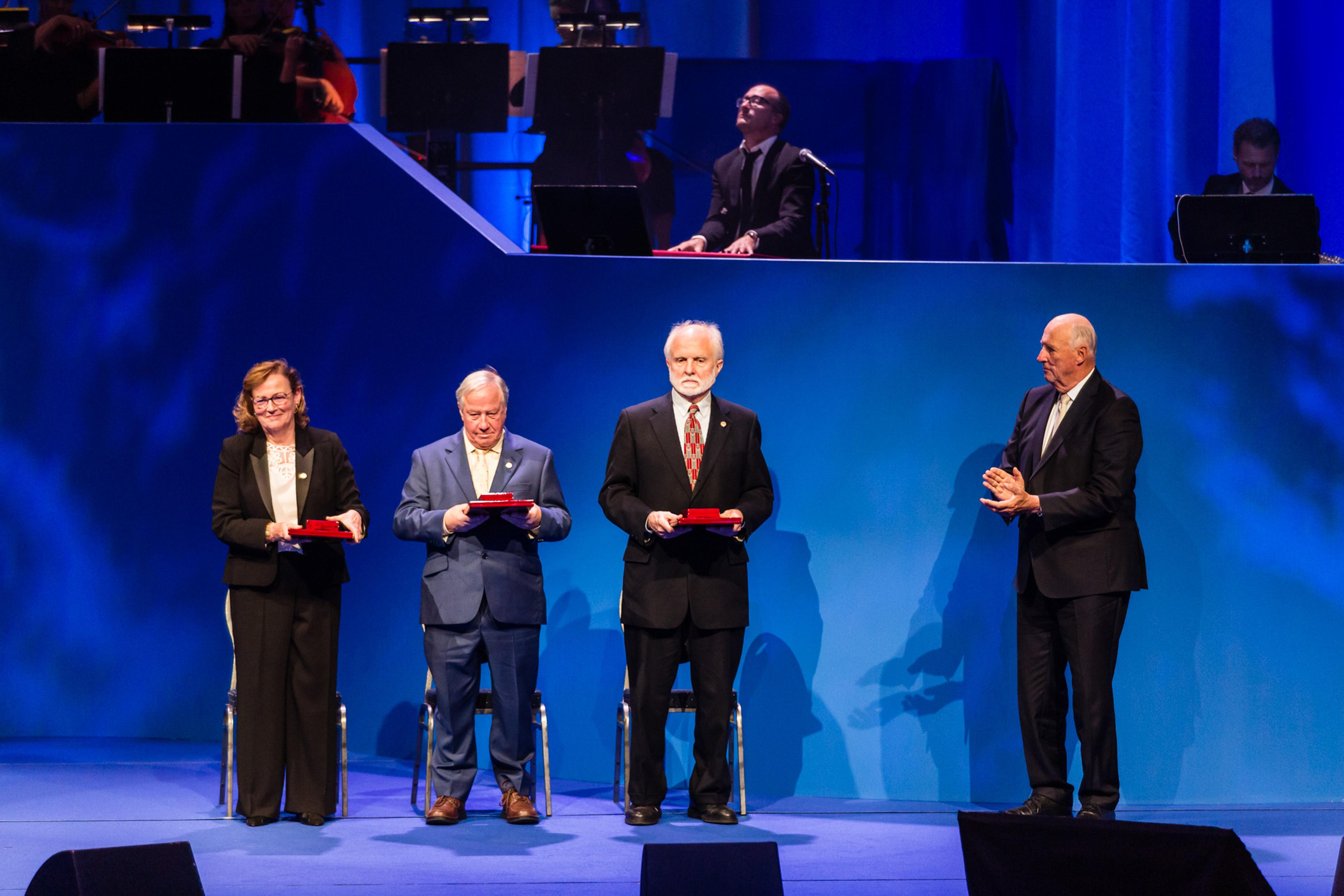
The 2018 Kavli Prize neuroscience laureates on stage in Oslo Concert Hall after having received the awards from His Royal Highness King Harald. From Left: Christine Petit, Robert Fettiplace, A. James Hudspeth and HRH King Harald. (Photo credit: Thomas Eckhoff)
By 1993, I had established my own laboratory at Institut Pasteur. Fascinated by the amazing performances of sensory perception, I decided to use a human genetic approach to investigate olfactory system development.Kallmann’s syndrome is the only hereditary syndrome known to cause complete anosmia, a lack of the sense of smell. We identified the first causal gene for Kallmann’s syndrome, KAL1 and contributed to the discovery of three others: FGFR1, PROK2, and PROKR2. We then showed that the protein encoded by KAL1, ANOSMIN-1, is an extracellular matrix glycoprotein involved in axon guidance and promoting the branching essential for the patterning of olfactory bulb output neuron projections onto the olfactory cortex.
Hearing
Through systematic explorations of the molecular mechanisms underlying the development and physiology of the other sensory systems, I came to realize, soon after discovering KAL1, that we actually knew next to nothing about the molecular physiology of the auditory system. Hearing is a sense intimately linked to the cognitive functions of human communication through language and music, and many concepts had already been developed concerning the principles of its functioning, mostly by physicists. So why was so little known about its functioning at the molecular level, even within the sensory organ, the cochlea? The answer lay in the very small number of each type of cochlear cell, incompatible with the resolution of the available biochemical and classical molecular genetic methods. Such constraints do not apply to the genetic approach. I thus opted for a neurogenetic dissection of the structure and function of the auditory system, focusing on the inherited forms of sensorineural deafness. However, several obstacles precluded a straightforward genetic approach. We overcame them by studying large consanguineous families affected by deafness and living in geographic isolation, mostly in North Africa and the Middle East. We thus mapped the first two loci for autosomal recessive hearing impairment, DFNB1 and DFNB2, to human chromosomes; a number of other laboratories subsequently initiated genetic studies of deafness using the same strategy. In these early days of disease-gene identification, with the human genome only partially sequenced, we developed an invaluable source of candidate genes by pulling out genes preferentially or specifically expressed in the cochlea. Wethus rapidly identified a number of genes responsible for severe-to-profound non-syndromic (isolated) or syndromic deafness, about 20 genes in all. Unsurprisingly, the first genes we identified were those responsible for the most frequent forms of deafness and almost all encoded previously unknown proteins. Most turned out to be components of the sensory hair cells of the inner ear. They included proteins of the hair bundle, the mechanoreceptive structure responding to sound stimulation, such as the PDZ domain-containing proteins HARMONIN, WHIRLIN, NHERF-1, NHERF-2 and another scaffolding protein, SANS, the transmembrane and membrane-associated extracellular proteins VEZATIN, PHR1 and STEREOCILIN, the unconventional myosin MYOSIN-VIIA and the kinociliary protein, CDC14A. They also included the hair-cell synapse proteins KCNQ4 and OTOFERLIN, the components of the tectorial membrane (a gel overlying and stimulating the hair bundle) OTOGELIN and OTOANCORIN, the major otoconial protein OTOCONIN-95, and a number of other proteins, including PEJVAKIN, EYA1, SIX1and AK2.
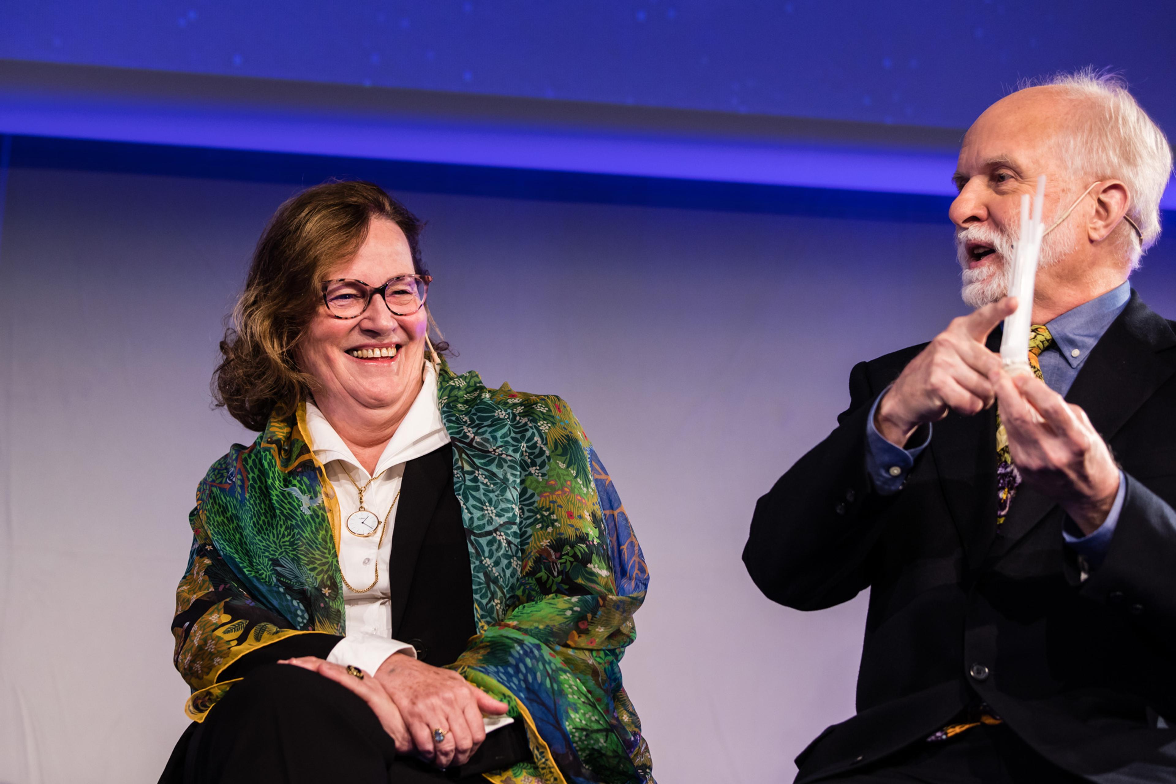
Christine Petit and A. James Hudspeth on stage during the interview session in the Kalvi Prize week in Oslo. (Photo credit: Thomas Eckhoff)
The physiological insight gained from audiometric tests in humans is too rudimentary to elucidate the function of these proteins, nevertheless an in vivo context is required to evaluate the interplay between the various sound-induced mechanical forces in cochlear sound processing. The cochlea is highly conserved in mice and humans. Engineered mouse models of human deafness, conditional knockout mice and mice carrying particularly informative missense mutations, analysed in details and combined with multidisciplinary studies, proved to be the most effective tools for exploring the molecular mechanisms of hearing based on “deafness genes”. To this end, scientists with complementary expertise, specifically in biochemistry, electrophysiology and biophysics joined the laboratory, and long-term collaborations were established with physicists, especially Paul Avan.

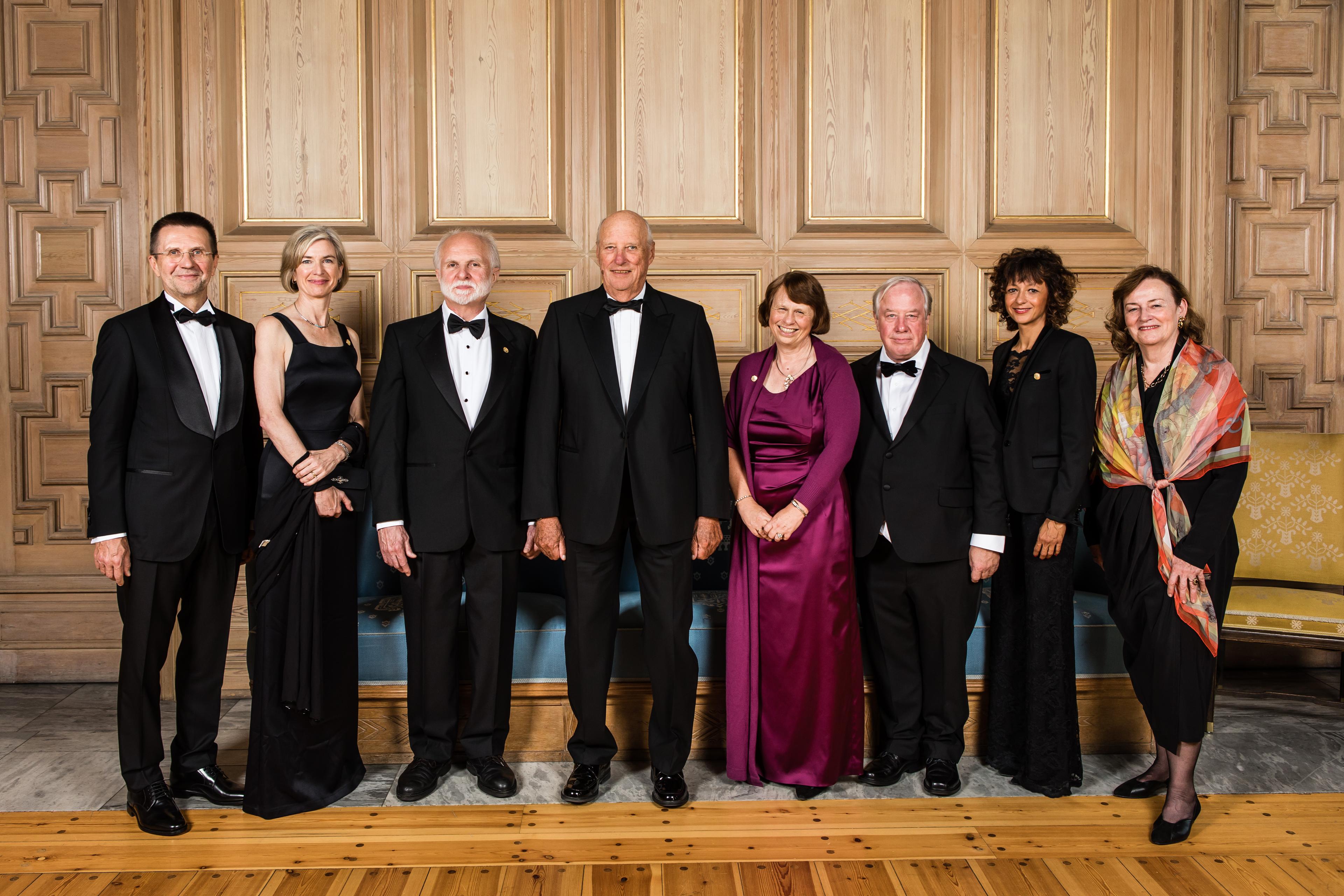
The 2018 laureates at the reception in the Munch room at Oslo City Hall, from left: Virginijus Šikšnys, Jennifer A. Doudna, A. James Hudspeth, His Royal Highness King Harald, Ewine van Dishoeck, Robert Fettiplace, Emmanuelle Charpentier and Christine Petit (Photo credit: Thomas Eckhoff).
Unsurprisingly, the identification of “deafness genes” revealed principally the molecular mechanisms underlying cochlea-specific functions. The frequent targets in hair cells are the hair bundle, a tuft of microvillus-like structures called stereocilia that processes sound and operates mechanoelectrical transduction (MET), hair cell-to-cell junctions which are particularly resilient to continuous sound-induced mechanical stress, and the hair cell synapses which display highly temporally precise, rapid and sustained neurotransmitter release.
Assuming that causal genes for syndromic forms of deafness encode proteins from the same network, we began deciphering cochlear protein complexes by studying the proteins encoded by genes defective in Usher syndrome (sensorineural deafness associated with blindness) type 1. We showed that MYOSIN-VIIA, HARMONIN, CADHERIN-23, and SANS form a complex anchoring the embryonic hair bundle lateral fibrous links and, subsequently, the tip links, to the actin filaments of stereocilia. We found that SANS is essential for tip-link maintenance and HARMONIN- b for preventing full tip-link relaxation. We demonstrated that protocadherin-15, which forms the basal part of tip-links, switches from several functionally redundant isoforms during embryogenesis to a single specific isoform in adults, revealing the existence of a process of MET machinery maturation. Our studies of inter- and intramolecular interactions showed how MYOSIN-VIIA, HARMONIN and SANS operate at the F-actin insertion point of the upper part of the tip-link, consisting of cadherin-23. We uncovered the presence of the Usher-1 adhesion protein complex in photoreceptors too, accounting for the retinitis pigmentosa observed in this syndrome. It forms an adhesion belt associated with calyceal processes, (neglected microvillus-like structures) involved in regulating disk and lamella sizes in photoreceptor outer segments.
Beyond this reductionist view, this approach captured a broader picture of integrated physiology. Mouse models of Usher-1 syndrome revealed relationships between the MET machinery and stereocilium size. Hair-bundle morphology regulates hair-bundle activity, and we thus showed the converse to be true.We found that the various fibrous links between hair-bundle stereocilia, which had previously been largely ignored, are essential for hair-bundle sound processing and morphogenesis. The lateral fibrous links of embryonic hair bundles are required for early stereociliary cohesion during hair-bundle development; the ankle links, composed of proteins encoded bycausal genes for Usher syndrome type 2, are essential for the development of the hair-bundle functional polarity. Our discovery of stereocilin, also encoded by a deafness gene, revealed the key role of the uppermost lateral links (the top connectors) and the crown of the outer hair-cell hair bundles in tectorial membrane attachment. By a further genetic dissection of the top-connectors and attachment links we recently showed these links to have a similar molecular composition, and therefore presumably also similar mechanical characteristics. The top-connectors ensure OHC stereociliary bundle cohesion, allowing the parallel gating of mechanotransduction channels required for the generation of large distortion products of oto-acoustic emissions (DPOAEs) in the ear canal. This touches on one of the many medical applications arising from our results. Audiometric tests frequently include DPOAE recordings, the interpretation of which has been clarified by these advances.
We also revealed the existence of a specific exocytotic molecular machinery at the first auditory system synapse, the inner hair-cell synapse, which is glutamatergic. This machinery includes OTOFERLIN, encoded by a deafness gene, a multi-C2-domain transmembrane protein of synaptic vesicles. Its synthesis begins when synaptotagmins 1 and 2 are no longer detected. We revealed a key role for otoferlin in synaptic release, as a Ca+2+ sensor for neurotransmitter release and synaptic vesicle replenishment in the active zone at the inner hair-cell ribbon synapse. Ferlins are considered to be ancestral to synaptotagmins. The similarity between the roles of otoferlin and synaptotagmins should prompt studies of interactions between otoferlin and other synapse components, including the synaptic SNARE complex, the v- and t-SNARE proteins of which remain unidentified.
Another facet of the auditory system was also brought to light by studies of “deafness genes”. We found that peroxisomes protect the auditory system against the deleterious effects of overexposure to noise, the major environmental cause of hearing loss. Loud sounds induce an adaptive proliferation of peroxisomes triggered by PEJVAKIN, we identified as a redox sensor encoded by a noise susceptibility gene.
Finally, we have recently begun to focus on developing gene therapy for preventing and curing hearing impairment, providing proof-of-concept for the efficacy of this approach in several mouse models of human deafness. However, all methods for hearing restoration acting on the peripheral auditory system are dependent on auditory cortex plasticity. It is generally assumed that defects of the peripheral auditory system are not associated with intrinsic defects of the central auditory system. Our recent findings challenge this view, by showing that the tip-link components cadherin-23 and protocadherin-15 are expressed by the precursors of a parvalbumin-positive interneuron population restricted to the auditory cortex, and demonstrating the requirement of these proteins for the embryonic migration of these cells. This finding raises many questions about the evolution of this sensory system and has major potential implications for patient management. Could this also be the start of the extension of genetic dissection to the central auditory system?
Teamwork
These advances are the fruit of real teamwork. They owe much to the excellence of my coworkers.
Family
I would like to thank my husband, Jacques, for our life together since our first year at university. I appreciate how lucky I have been, thanks to him, to have been able to enjoy both a highly active professional life and a happy family life, with a daughter who is now a doctor, a son who is a jazz cellist, and many long-standing friends with whom we have shared a great deal.