


Keep your door open to new ideas!
As told by Harry Orr
The path that brought me to this exciting and rewarding point is blessed with many stellar mentors, colleagues, trainees, family, and friends, in and out of science. I was born on May 9, 1949, in Clifton Forge, a small town in the western mountains of Virginia, USA. My father worked as a civil engineer for the C&O Railroad and we relocated with each of his promotions. Our final move was to Birmingham, Michigan outside of Detroit. In 1962, just before we were to move to West Virginia, my father passed away from a heart attack. My mother decided to keep our family (herself and four children) in Michigan. She worked as a medical technologist in a hospital laboratory. My fondness for biology, which I discovered when I took biology in high school, most likely comes from her influence.

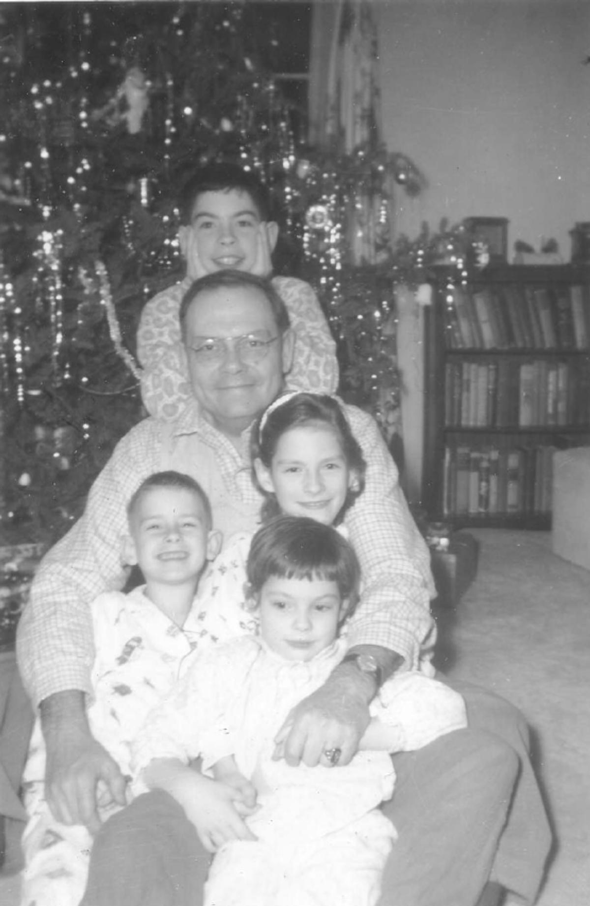
Me (on top), my dad and my three siblings, 1961
As an undergraduate biology major at Oakland University in Rochester, Michigan I had the great fortune of being a teaching assistant for freshman biology and working, most importantly, as a research assistant in the NIH-funded laboratory of Dr. Michael V. Riley, an outstanding mentor. In Mike’s lab, I gained a fascination for lab bench research while working on the bovine corneal endothelium. It was there that I published my first paper, and it was Mike who suggested I apply to a relatively new neuroscience graduate program at Washington University in St. Louis.
At Washington University, my Ph.D. thesis examined the neurochemistry of vertebrate micro-dissected retinal layers. Though the bulk of my research was performed in the laboratory of Dr. Oliver (Ollie) H. Lowry in the Department of Pharmacology, I was co-advised by Adolph I. Cohen of the Department of Ophthalmology and James A. Ferrendelli Departments of Neurology and Pharmacology. The Lowry lab was very skilled in performing sensitive biochemical assays on micro-dissected tissue samples, ideal for studying the regional specificity of biochemical properties of the retina. Drs. Ferrendelli and Cohen provided strong expertise in measuring levels of cyclic nucleotides, cAMP and cGMP, in relation to neuronal function/dysfunction and retinal biology, respectively. In my opinion, the capstone of my thesis was the finding that cGMP is highly concentrated in retinal photoreceptors and undergoes substantial increase in the portion of the retina containing photoreceptor synapses with dark adaptation. This work formed the basis of subsequent studies showing that cGMP is central to excitation of vertebrate retinal rod photoreceptor cells. Sodium channels in the plasma membrane of rod outer segments are kept open in the dark by a high level of cGMP. Light closes these channels by activating an enzymatic cascade that leads to the rapid hydrolysis of cGMP. Looking back, I believe my graduate training showed me how much I enjoyed studying the molecular pathways that underlie physiological processes.
“Looking back, I believe my graduate training showed me how much I enjoyed studying the molecular pathways that underlie physiological processes.”
Harvard University
Ollie’s mentoring had another major impact on my career when he suggested I learn the new molecular genetic approaches that were being developed and suggested a postdoctoral position with a colleague of his at Harvard University might be an excellent choice. In 1976, with a Ph.D. in neuroscience, I joined the laboratory of Dr. Jack L. Strominger in the department of biochemistry and molecular biology, The Biological Laboratories, Harvard University, Cambridge, Massachusetts. In Jack’s lab, I worked on defining key molecular features of the highly polymorphic membrane proteins encoded by the human major histocompatibility complex, i.e., Class I the HLA antigens. Besides being target antigens during graft rejection, these proteins are important in the immune system’s recognition of virally infected cells and tumor cells. By generating analyzing protein amino acid sequence data from HLA antigens, I participated in studies that showed a structural and evolutionary relationship between HLA Class I antigens and immunoglobulins as well as providing insight into polymorphic regions important for regulation of the immune response by HLA antigens.

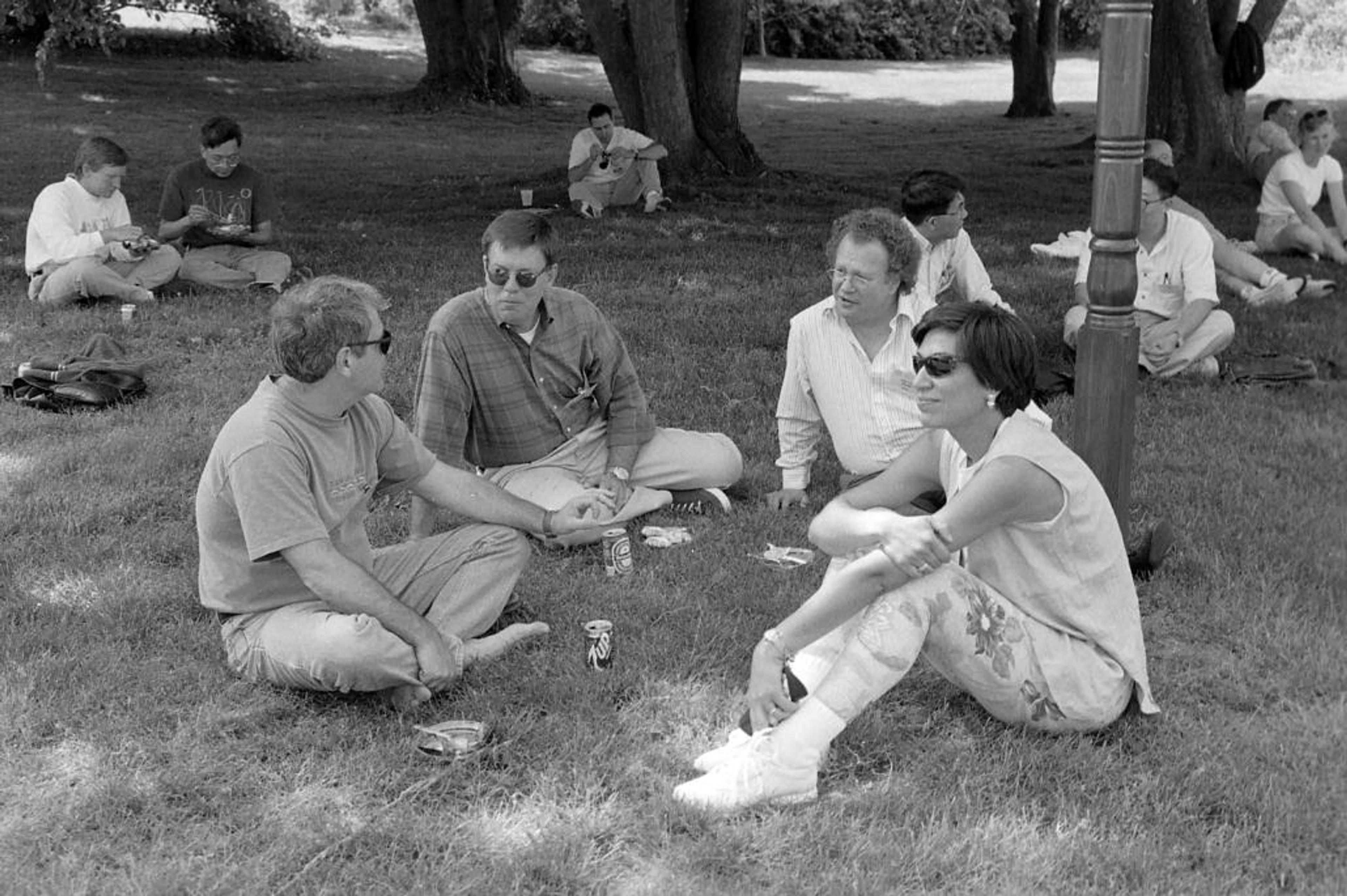
Me, Steve Warren, Jean-Louis Mandel and Huda at the 1996 Cold Spring Harbor neurodegenerative disease meeting.
I was fortunate to work with Hidde L. Ploegh, a graduate student from the University of Leiden in the Strominger group, on generating the first HLA-specific cDNA clone. Importantly, this HLA cDNA clone corresponded to a region highly conserved among HLA Class I antigens.
Back to the Midwest
In 1980, I attended a Keystone Immunology meeting. On a bus ride from the conference site to the airport for my flight home I happened to sit next to David Zarling from the University of Minnesota, who informed me that the university was recruiting immunology faculty. Eager to return to the Midwest, I followed up on David’s information.
In 1981, I accepted an independent faculty position as a member of the Immunobiology Research Center in the Department of Laboratory Medicine and Pathology at the University of Minnesota Medical School in Minneapolis. There, I embarked upon analyzing the genomic organization of HLA Class I genes. Key to the success of these studies was my collaboration with Dr. Robert DeMars from the Laboratory of Genetics and Department of Human Oncology, University of Wisconsin, Madison, Wisconsin. Bob developed a very elegant approach to generate γ-radiation-induced HLA variants from a human lymphoblastoid cell line. The HLA cDNA clone was used to correlate specific restriction fragments with expression of specific HLA alleles in the γ-radiation-induced HLA variants. This approach quickly led to the finding that the HLA Class I genes are a multi-gene family with some genes mapping distal to the HLA-A locus that was previously thought to be the most telomeric HLA Class I gene. One prominent outcome was identification of the HLA-G class I gene, which others subsequently showed is important the establishment of maternal-fetal tolerance.
Long-standing “brothers”
In the summer of 1983, I presented our HLA gene mapping work at the conference on Somatic Cell Genetics Federation of American Societies for Experimental Biology at the Vermont Academy. Many prominent human and molecular geneticists attended and were using similar approaches to determine the genomic position of genes in which mutations caused diseases. I was fortunate to meet and befriend three other junior investigators Steve Warren, David Nelson, and Web Cavenee, who were using similar approaches to map and isolate disease genes (Fragile X syndrome and tumor suppressor genes in retinoblastoma). All three became colleagues with Steve and David becoming my long-standing “brothers” in human genetics as involved members of the American Society of Human Genetics.
Ataxia proposition
As a junior faculty member at the University of Minnesota, my group continued the genomic/molecular genetic analyses of HLA Class I genes when one day in 1984, Dr. Larry Schut walked into my office. Larry, a neurologist at the University of Minnesota, came from a large family affected by a genetic form of the neurodegenerative disease ataxia. He, along with Dr. V. Elving Anderson and Jonathan L. Haines (then a graduate student), had just demonstrated that the ataxia gene in Larry’s family was genetically linked to the HLA complex. Given my neuroscience background and expertise in using HLA DNA probes, Larry thought the group might be interested in using our HLA probes to further map and eventually isolate the ataxia gene in his family. We jumped at the opportunity to embark upon a path of ataxia research. To this day, I am grateful that Larry walked into my office with his ataxia proposition. I often tell this story to trainees as an illustration of why it is important in one’s research career to keep your door open to new ideas and directions.
“I often tell this story to trainees as an illustration of why it is important in one’s research career to keep your door open to new ideas and directions.”
The Zoghbi-Orr collaboration
In collaboration with my colleague Dr. Stephen S. Rich, a highly skilled computational geneticist, we first localized the ataxia gene to a rather broad region several centimorgans telomeric of HLA-A using our HLA DNA probes and other newly identified DNA markers from the short arm of chromosome 6 (6p). I mentioned our new ataxia gene cloning project to Dr. Arthur Beaudet from Baylor College of Medicine during his visit to the University of Minnesota to give a seminar on his work with cystic fibrosis. Art quickly responded that I should contact an outstanding trainee at Baylor who was also working on the mapping and cloning of an ataxia gene from chromosome 6p, Dr. Huda Zoghbi. When I contacted Huda, she informed me that their findings indicated the ataxia gene they were hunting was located centromeric to HLA. In addition, Huda very graciously shared with me the use radiation hybrids, an approached developed by Dr. David Cox, as a means to map and isolate additional chromosome 6p DNA. Huda later shared with us a newly isolated highly polymorphic DNA marker, D6S89, from chromosome 6p. Using this marker and additional polymorphic probes, Dr. Laura Ranum (a postdoc), Lisa Duvick (a research technician), and Ming-yi Chung (a graduate student), obtained data that strongly indicted the ataxia gene was positioned in a defined region telomeric to HLA-A. Prior to the publication of our refined localization of the ataxia gene, I received a call from Huda telling me that the ataxia gene in the family she was studying was also telomeric to HLA-A, and our two groups were going after the same gene. Huda proposed that we collaborate, and after a pause I agreed. Clearly, this was the smartest decision I have ever made in my scientific career.
“Huda proposed that we collaborate, and after a pause I agreed. Clearly, this was the smartest decision I have ever made in my scientific career.”
The Zoghbi-Orr collaboration localized the ataxia gene to 1 x 10⁶ base pair region on chromosome 6p and cloned this region on a yeast artificial chromosome (YAC) contig. Previous work showed the presence of anticipation, i.e., the disease progressively became more severe as the gene passed onto successive generations in the Schut family.

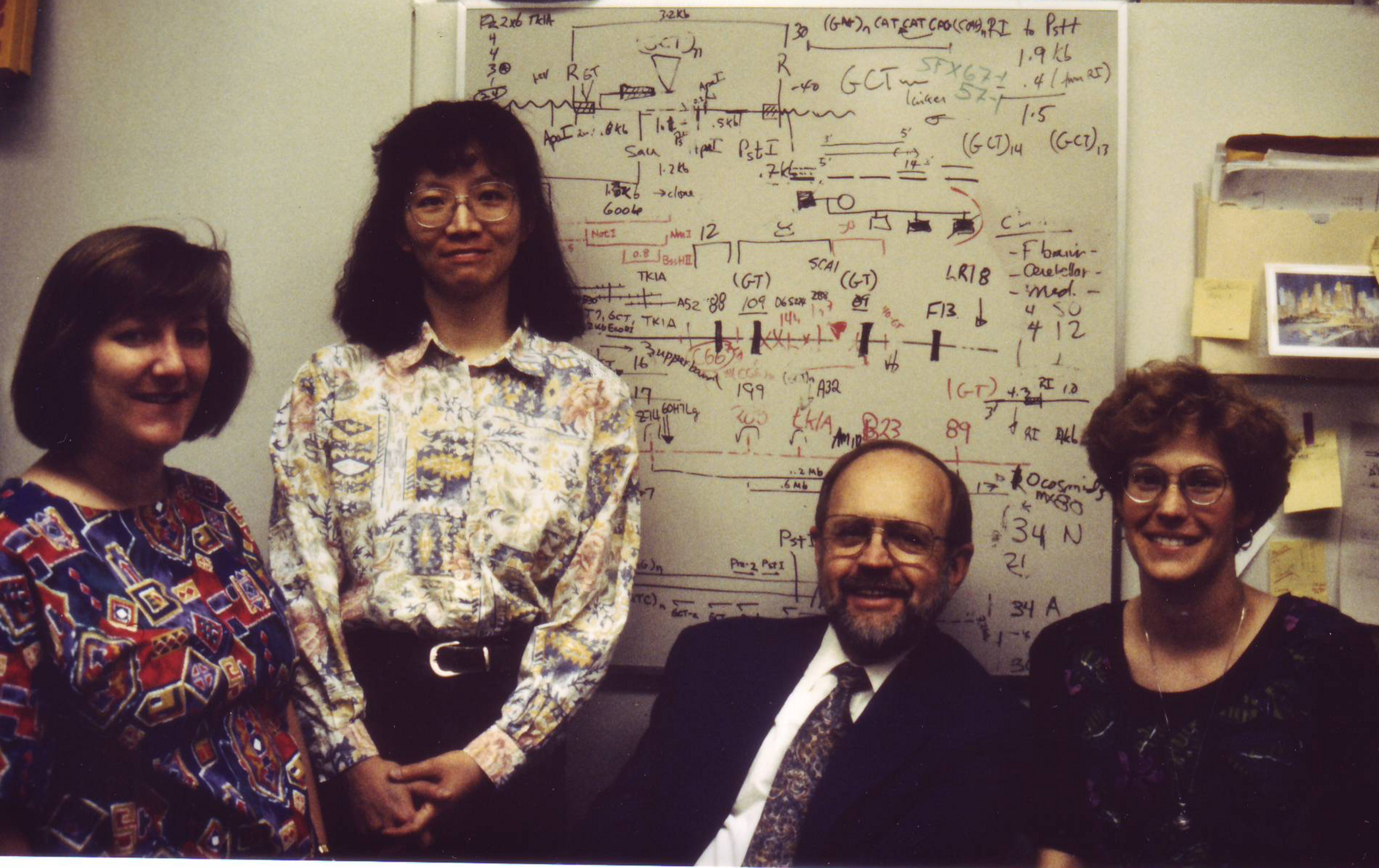
The SCA1 genomics group 1993; Lisa Duvick, Ming-yi Chung, Larry Schut, and Laura Ranum
Importantly, it was becoming apparent from work on other unstable repeat-based neurological diseases that anticipation could be explained by the expansion of an unstable trinucleotide repeat in the affected gene. This finding guided us identifying a CAG repeat in our own ataxia YAC contig. This was accomplished using a single copy segment of DNA adjacent to the CAG repeat to probe a Southern blot of a family where the father had a late onset disease, and the affected offspring had a juvenile onset of ataxia. We reasoned if the ataxia gene was due to expansion of a CAG repeat it would be evident on Southern blots of this family having prominent anticipation. In 1993, the two groups simultaneously found that ataxia in the families under study was due expansion of the CAG repeat in chromosome 6p YAC contig. We first announced the cloning and characterization of the ataxia gene at a meeting on Capri, Italy that summer. With the cloning and characterization of the ataxia gene linked to the HLA complex the disease was designated as spinocerebellar ataxia type 1(SCA1). The gene and protein affected in SCA1 were designated Ataxin-1 and ATAXIN-1, respectively. In Ataxin-1, the CAG encodes a polyglutamine tract within the protein, placing SCA1 among the group of polyglutamine neurodegenerative diseases.
With the cloning of the SCA1 gene, Huda and I planned how best to divide the next series of experiments. Our focus was to generate a transgenic mouse model that would allow us to study the cerebellar Purkinje cell aspects of SCA1 pathogenesis. Due to the talented efforts of two individuals, Dan Nordquist and Sylvie Vandaele, the regulatory region from the Purkinje cell-specific gene Pcp2 was identified and shown to effectively direct transgene expression to Purkinje cell in mice. Lead by Eric Burright, a postdoc, we generated and described transgenic expressing ATAXIN-1 with an expanded polyglutamine tract that nicely replicate SCA1-like Purkinje cell pathologies and subsequent deficits in motor performance. Over the years, and through the efforts of talented postdocs, graduate students, and technicians, we used this and variations of this SCA1 Purkinje cell mouse model to identify a molecular pathway that underlies Purkinje cell disease. Importantly, these studies consistently show it is an expression of the ATAXIN-1 protein with an expanded polygutamine tract driving Purkinje cell pathogenesis. In addition to the polyglutamine stretch, features of the ATAXIN-1 protein we found to be critical for disease are:
1) Phosphorylation of ATAXIN-1 at serine 776, which regulates its interaction with the chaperone 14-3-3 and stability of ATAXIN-1;
2) Entry of ATAXIN-1 into Purkinje cell nuclei is critical for Purkinje cell pathogenesis. A single amino acid mutation in the nuclear localization sequence of ATAXIN-1 prevents its entry into Purkinje cell nuclei and development of disease; and
3) The AXH domain of ATAXIN-1 (residues 560 to 690) is critical for the interaction of ATAXIN-1 with the transcriptional repressor Capicua (Cic). Mutations in the AXH that block the ATAXIN-1/Cic interaction prevent Purkinje cell disease.
Using a conditional Pcp2- ATAXIN-1 transgenic mouse model in which expression of the transgene could be regulated by administration of a drug, we found that the earlier expression of the mutant ATAXIN-1 transgene was blocked the better mice recovered. However, if the transgene was stopped at a late stage of disease remaining Purkinje cells have some ability to repair damage caused by mutant ATAXIN-1. Intriguingly, we also showed if expression of the mutant ATAXIN-1 transgene was delayed until development of the cerebellum was complete (in mice, three weeks postnatal) this led to a substantial reduction in severity of Purkinje cell disease suggesting that an aspect of Purkinje cell disease is due to compromising development of these neurons by mutant ATAXIN-1. In terms of potential treatments for SCA1, we used our SCA1 mouse models to demonstrate the therapeutic efficacy of using antisense oligonucleotides targeting the reduction ATAXIN-1 RNA. Recently, we reported that activation of the cholecystokinin 1 receptor on Purkinje cells of SCA1 and SCA2 transgenic mice is neuroprotective.
Current efforts of our group focus on challenges in understanding pathogenesis of SCA1, and neurodegenerative diseases in general, as well as elucidating the relative contribution of each molecular pathway altered has in pathogenesis in the different regions of the brain affected. To this end, we are performing a molecular characterization of the importance of nuclear entry of mutant ATAXIN-1 in regions/cell types other than cerebellar Purkinje cells. In this work we are using CRISPR-Cas to mutate the NLS of mutant ATAXIN-1 in a SCA1 knockin mouse. Using a recently developed floxed ATXN1[146Q] mouse model we will determine the regional/tissue contributions to SCA1-ilke phenotypes. Finally, we continue to examine the extent to which manipulation of the Cholecystokinin - Cholecystokinin 1 receptor pathway can serve as a potential therapeutic target for treatment of SCA1, as well as other SCAs that involve Purkinje neuron degeneration and perhaps neurons in other SCA affected brain regions.

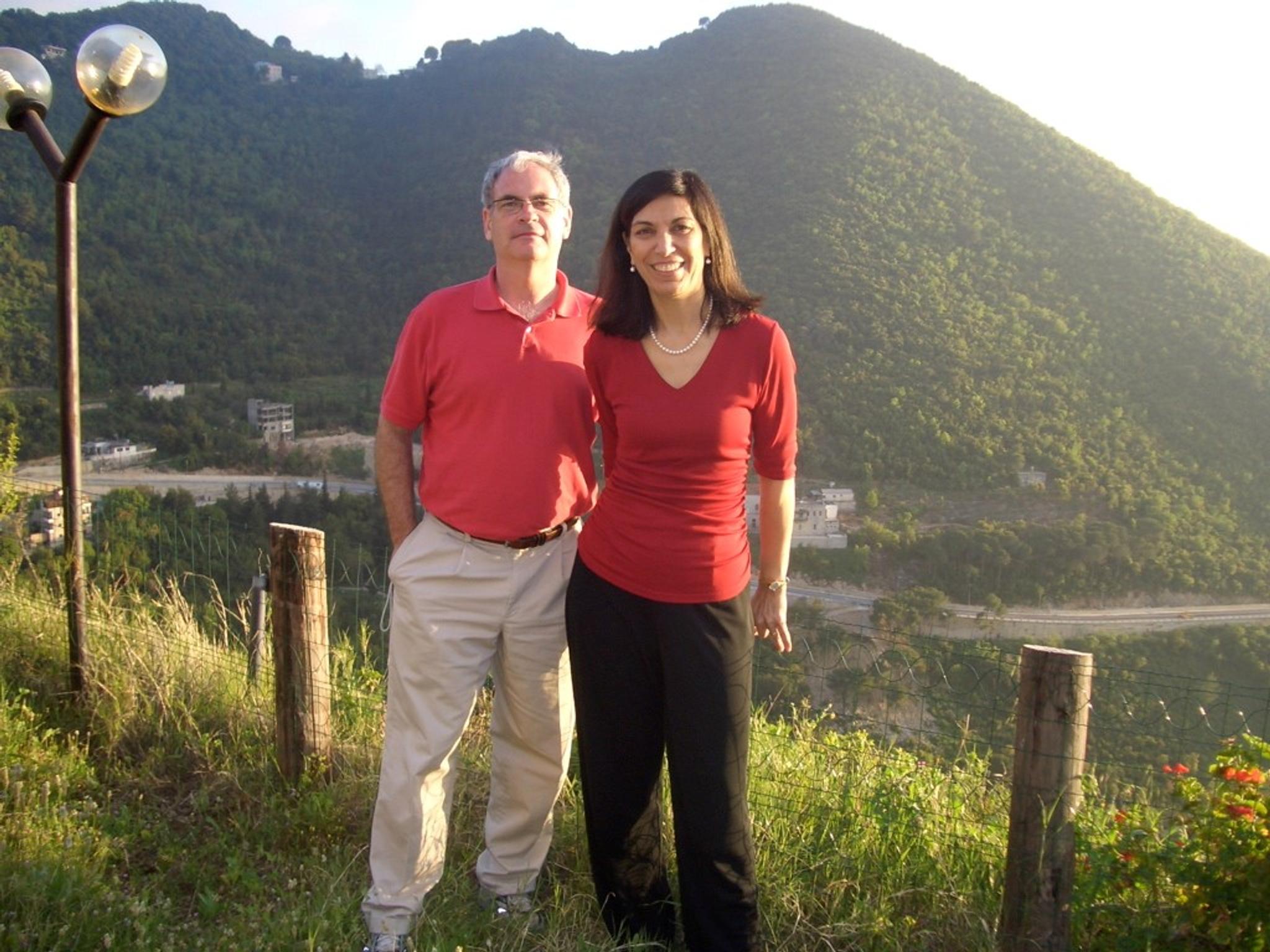
Me with Huda in Lebanon, 2009
Collaboration and friendship
I continue to enjoy my life as a human geneticist/neuroscientist. My long-standing and ongoing collaboration and friendship with Huda is a shining example of how one can benefit from the collaborative aspect of research. Because of this relationship, I had the good fortune of traveling to Beirut in April 2009 and meeting Huda’s family and seeing where she grew up. I found Beirut to be very beautiful and the Lebanese to be extremely open and friendly. At the birth of my younger daughter Olivia, Huda sent us a collection of children’s books. I am pleased to say this tradition continues. When Huda’s daughter Roula gave birth, my wife and I sent them a similar assortment of books.
Banner year
The year 1993 was a banner year for me both personally and professionally, as I married my wife Bonnie in the same year that we cloned the ATXN1 gene. These two events had an immeasurable impact on the path of my life, and I am a better man for it. Bonnie is the love of my life, life-partner, and mother of our daughter, Olivia. Bonnie’s support and counsel over the years has helped me keep a positive perspective, critical for both my personal and scientific lives.

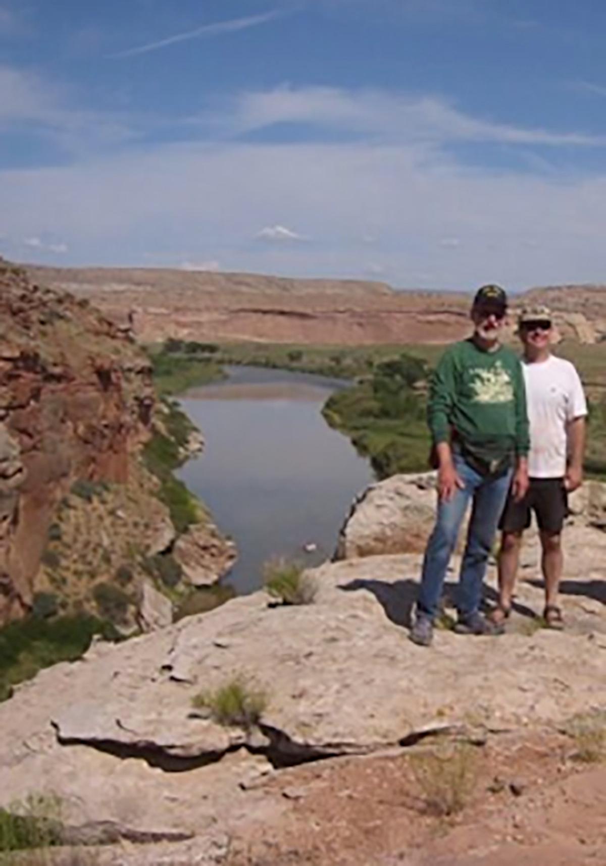
University of Minnesota colleague Dick Poppele and me on the Westwater overlook of the Colorado River.
Work-life balance
As a postdoc at Harvard, Jack Strominger advised me of the importance of work-life balance, as well as taking breaks for adventures. Following this advice, for the past 30 years I have taken an annual family vacation with kids and now grandsons to Mackinaw Island in northern Lake Huron. No cars are permitted on the island, making bike riding very relaxing.
In addition, I take an annual white water rafting trip with friends and guided by Brenda of Sheri Griffith Expeditions out of Moab, Utah on either the Green, Yampa, or Colorado rivers. On these trips I am off the grid and immersed in fantastic scenery, while spending quality time with old and new friends. The amazing archaeology, which reminds me that others have gone before, in this case the ancient Puebloans.
I am very grateful to the many technical staff, students, postdocs, and collaborators for working with me to understand the importance of genetics in immunology and neurodegenerative disease. Key to the success of the group is Lisa Duvick the lab Senior Research Manager. Since Lisa joined the group on her birthday in 1986, she has played a critical role in all the accomplishments the research group have enjoyed over these years.


Family on Mackinaw Island, 2019. My two daughters Olivia and, son-in-law Tom, two grandsons Bennett and Jack, and my wife Bonnie