


2012 kavli prize in Neuroscience
2012 Kavli
Prize in
Neuroscience
The Norwegian Academy of Science and Letters has decided to award the 2012 Kavli Prize in Neuroscience to
Cornelia Bargmann, Winfried Denk and Ann Martin Graybiel.


"For elucidating basic neuronal mechanisms underlying perception and decision."
Committee Members
- Jon Storm-Mathisen (Chair), University of Oslo, Norway
- Stanislas Dehaene, INSERM-CEA, France
- Lily Jan, University of California, San Francisco, USA
- Bert Sakmann, Max Planck Institute of Neurobiology, Germany
- Charles F. Stevens, Salk Institute, USA
Citation from the Committee
Understanding how the brain receives information from the environment and processes it to make decisions is a major challenge in neuroscience. The prize winners have addressed this question in different organisms, from worms to primates. They have used a wide range of approaches and have invented new technologies.
Cornelia Bargmann has pioneered the study of how genetic programs control the operation of neural circuits. By exploiting the simplicity of the C. elegans nervous system, Bargmann has identified fundamental principles of neural circuit logic. The functional circuits involve fast synaptic transmission and volume transmission via amines and peptides as well as gap junctions – much like our nervous system – but are more amenable to genetic analysis. She provided the first evidence, in any animal, for the detailed neuronal pathway between a specific sensory receptor protein and behavior.
Winfried Denk has devised two experimental innovations of broad impact to studies of how individual neurons respond to synaptic inputs. He developed multi-photon microscopy, which revolutionized high resolution imaging in the living brain. Using this technique, he discovered that direction selectivity is computed locally in individual dendritic branches of starburst amacrine cells in the retina. His invention of serial block-face scanning electron microscopy then revealed the wiring asymmetry between these inter-neurons and the retinal ganglion cells that convey motion information to the brain.
Ann Graybiel has unravelled the modular architecture of the striatum, and discovered striatal plasticity underlying habit learning. Her detailed analysis of how multiple cortico-striatal loops change when animals learn new skills reveals the way neuronal circuits organize familiar motor patterns into action sequences.
The cortico-striatal associative loops are also important for working memory and cognitive control, and likely contribute to the mechanism of disorders such as schizophrenia, Parkinson’s disease, and addiction.
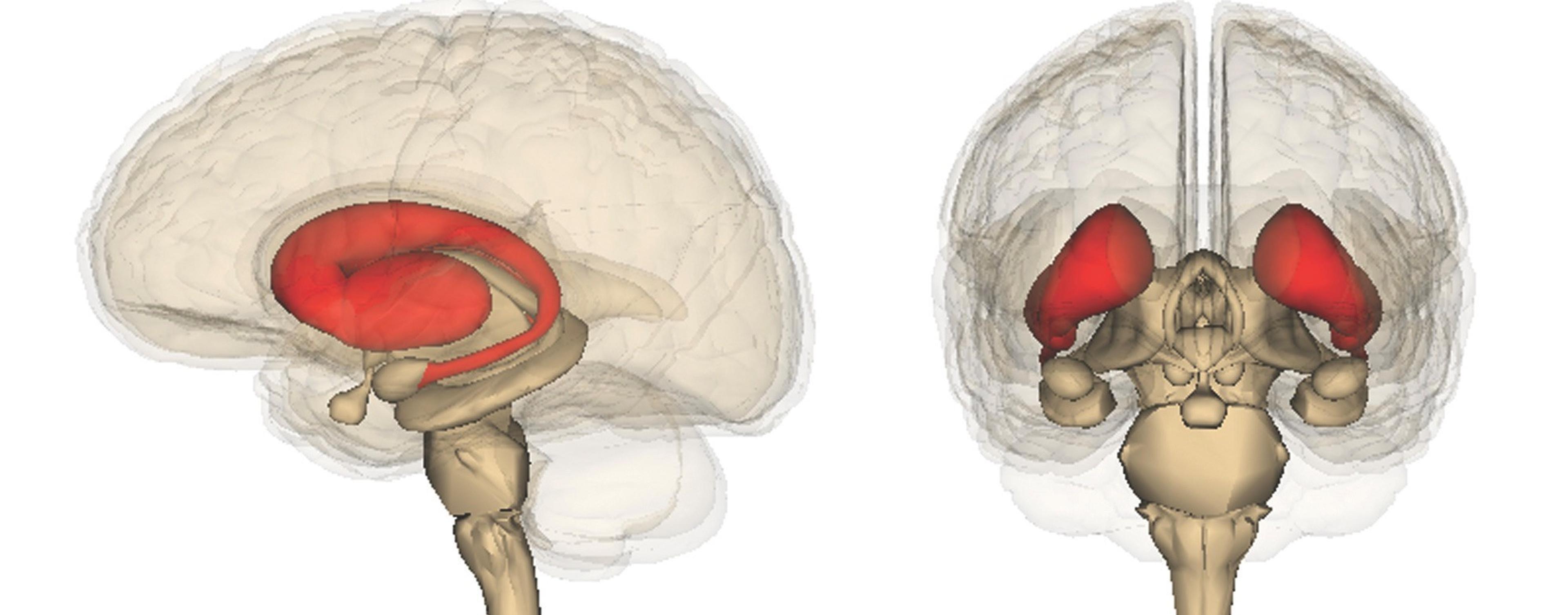
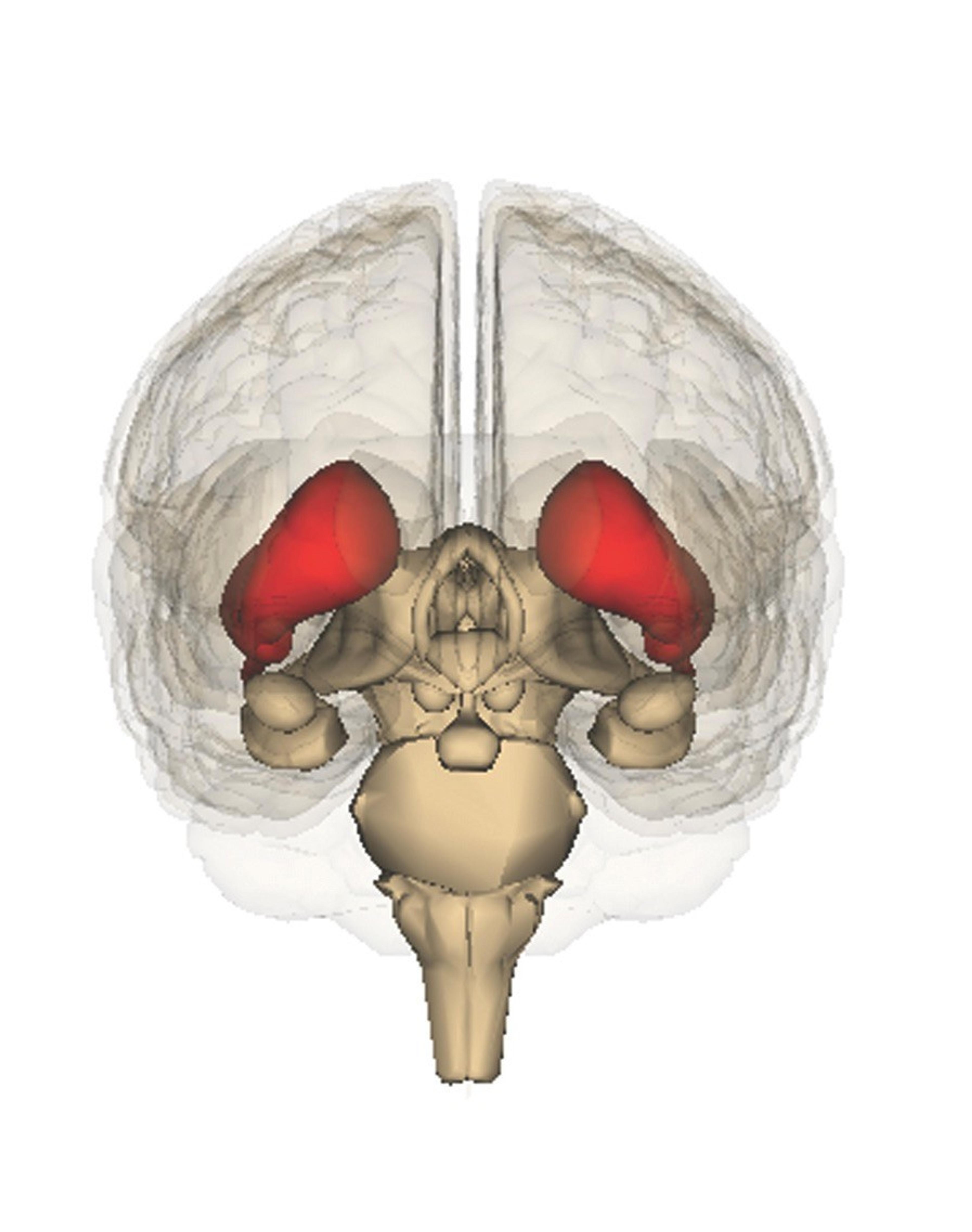
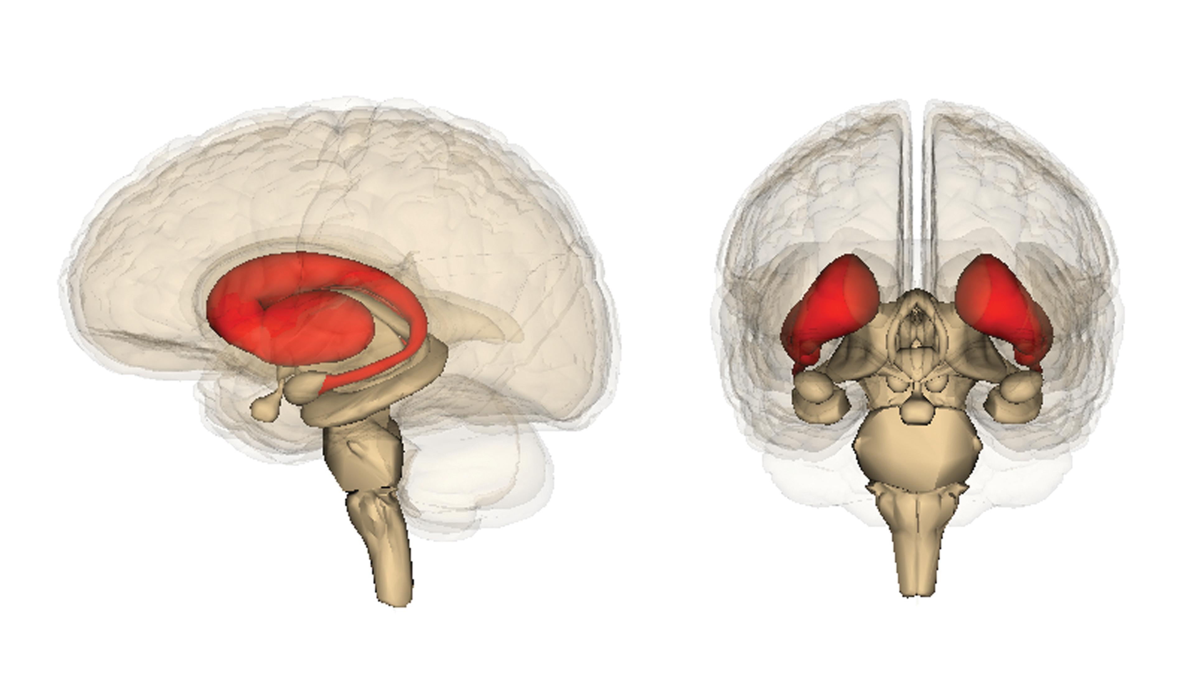
The striatum (red) plays an important role in the learning of new tasks by a ‘use it or lose it’ process of reinforcement of nerve signalling pathways. Abnormalities in the same neural circuits are linked to disorders such as Huntington’s disease, Parkinson’s disease, obsessive-compulsive disorder, and autism. (Photo credit: Anatomography, maintained by Life Science Databases(LSDB), under Creative Commons Attribute)
The 2012 Neuroscience Kavli Prize explained
The 2012 Kavli Prize for Neuroscience goes to three neuroscientists, Cornelia Bargmann, Winfried Denk, and Ann M. Graybiel, who have pioneered the study of how sensory signals pass from the point of sensation – whether the eye, the foot, or the nose – to the brain, and how decisions are made to respond. Each working on different parts of the brain, and using different techniques and models, they have combined precise neuroanatomy with sophisticated functional studies to gain a rounded understanding of their chosen systems.
By Julie Clayton
Using such terms as “pirouette,” “reversal,” and “deep omega turns,” Cornelia Bargmann, joint winner of the Kavli Prize for Neuroscience 2012, could be writing a review of a dance performance. And in a way, she is. But rather than people, she is describing the movements of tiny transparent worms as they wriggle about in laboratory dishes at the Rockefeller University in New York, where she holds the position of Torsten N. Wiesel Professor. Professor Bargmann has devoted more than two decades to the study of the nematode worm Caenorhabditis elegans (C. elegans) as a model organism for understanding how genes and the environment influence animal behavior.
A person tasting poisonous leaves or rotten meat may quickly spit out the food on account of its unpleasant taste, and learns from the experience to avoid such food in future. A child putting its hand in hot water will rapidly feel pain and withdraw the hand. But trying to understand the nerve signalling pathways involved is a gargantuan task, given the complexity of the human nervous system, with its billions of neurons and trillions of connections between them.

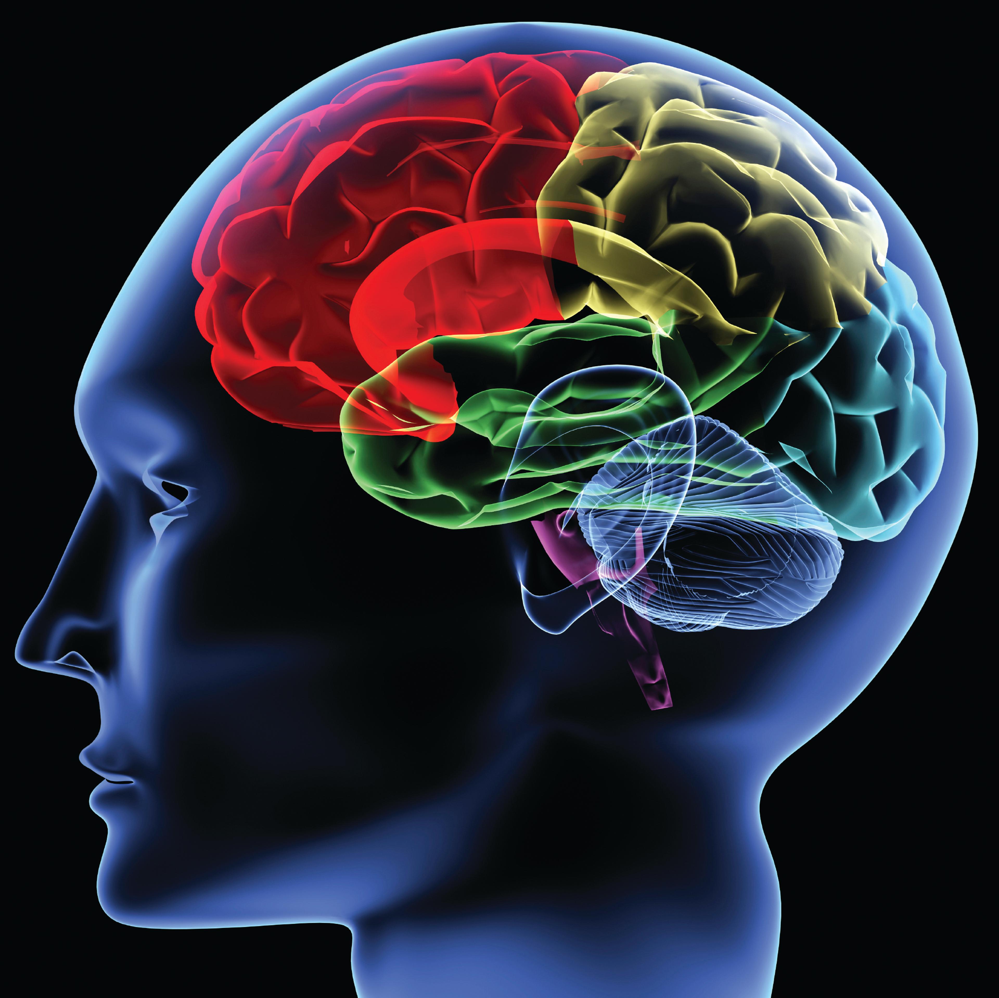
The brain’s time-keepers: Neurons in the forebrain and mid-brain fire rhythmically at different time intervals to help us coordinate our movements and behaviour. (Photo credit: Christine Daniloff/ MIT News Office)
Bargmann chose C. elegans. Just one millimeter long, and with a mere 302 neurons, this simple model organism was easy to grow and manipulate in the laboratory. Because each neuron has reproducible functions and connections, Professor Bargmann could define key aspects of neuronal development and function without the added complexity of studying mammals.
In her Howard Hughes Medical Institute profile, Professor Bargmann says, “When we understand the worm’s brain, we’ll be in a much better position to understand the complex functions of the human brain.”
Seeking out pleasant odors, learning to discriminate and avoid bitter tasting food, and choosing to group together or move apart – these are all examples of behaviors shared between C. elegans and more complex animals, and which are influenced by genetics, experience, and sensations. C. elegans depends heavily on its sense of smell to get around and find food.
Professor Bargmann’s pioneering work has provided important insights into the molecular switches and controls for C. elegans movement and behavior – many of which have human counterparts. She has investigated which neurons in C. elegans relay sensory information to interneurons and motor neurons, and defined key molecular signals and the genes encoding them.

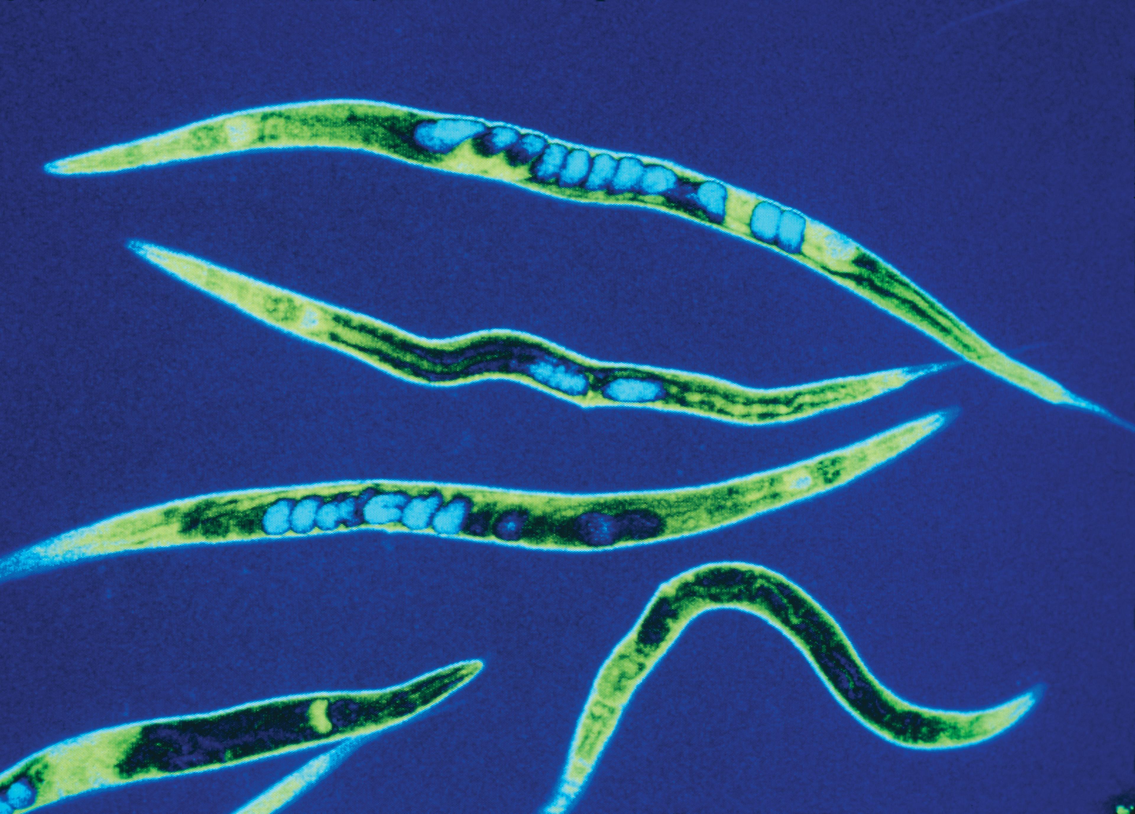
False-colour scanning optical micrograph of Caenorhabditis elegans (C. elegans). Cornelia Bargmann has for more than two decades used these millimetre-sized transparent worms as a model organism for understanding how genes and the environment influence animal behaviour. (Photo credit: James King-Holmes/Science Photo Library)
Studies published in 1997, for example, focused on the reflex-like response of C. elegans of moving toward, or away from, a particular odor, depending on whether it was attractive or repulsive. C. elegans has the ability to distinguish between hundreds of different odors due to a range of molecules – odorant receptors – on the surface of a cluster of neurons at the tip of its head. Bargmann provided the first evidence showing that the type of odor response in C. elegans – attraction or repulsion - was governed not by the specific odorant receptor, but by the neuron expressing it. By genetic manipulation, Bargmann and her team swapped the receptors for two different odorants between two neurons, and produced opposite effects to those that normally occur – a receptor for an attractive odor, now on a neuron that usually responded to noxious odors, caused worms to move away from the attractive odor source. And a noxious odor sparked movement toward it. This was the first evidence that neurons rather than the receptors determine odorant responses.
More recently, Professor Bargmann has taken chemotaxis studies to new levels with the development of an easy to replicate microfluidic system with which to quantify movement in both time and space in response to attractive odors.
With similar precision, Professor Bargmann has also identified many of the intracellular signalling pathways in C. elegans that relay information from cell surface odorant receptors (G protein-coupled receptors) to the interior of each sensory neuron. Her team has revealed some of the neurons and the receptors, neurotransmitters, and neuropeptides that act when the worms learn and adapt their behaviour after experience. These include the neurotransmitter serotonin, which is important when worms learn to avoid eating pathogenic bacteria.
Professor Bargmann was also intrigued by the observation that some worms tended to forage alone while others preferred to clump together, and suspected a genetic explanation. In 1998, she discovered that worms showing different foraging behavior expressed different variants of the neuropeptide receptor npr-1, which is closely related to the human neuropeptide Y receptor family, involved in appetite regulation, obesity, and anxiety. In 2002, she identified another four genes that promote social feeding and which are found on neurons responding to stressful stimuli such as pain and noxious chemicals. And in 2004, she added to these the effects of a haem-containing protein that detects changes in oxygen concentrations and directs worms to group together when surrounding oxygen concentrations are high.
“This is the first and probably only example of where the whole problem is worked out over the entire circuit from how the sensory receptor protein detects say an odor or pheromone to how the animal behaves,” says Professor Lily Jan, Investigator at the University of California San Francisco, and member of the Kavli 2012 Prize Committee for Neuroscience.
Winfried Denk, Director of the Max Planck Institute for Medical Research, Department of Biomedical Optics, in Heidelberg, Germany, has made a unique contribution to neuroscience by inventing two new experimental approaches that have transformed not only his own research but also that of other neuroscientists. The first invention was two-photon fluorescence microscopy and the second, serial block-face electron microscopy. Taken together, these two developments have enabled Professor Denk to crack a major puzzle in neurobiology: how mammals – including humans – see and detect motion. Scientists had been pursuing this by studying the retina (the light-sensitive layer of the eye) of rabbits and other experimental animals for almost 50 years and it had, in Denk’s words, “defied comprehensive explanation.”
The retina has multiple layers of cells beginning with photoreceptors (rods and cones), which respond to light and signal other cells in the retina. The signals are relayed, via bipolar cells and other interneurons including starburst amacrine cells, to retinal ganglion cells, which then project to the brain. An early finding was that some retinal ganglion cells are "directionally selective" – in other words, they only respond when a visual cue, such as a bright spot or a dark shadow, goes in one direction, and not when the light or shadow moves in the opposite (null) direction. For the rabbit, this could make all the difference in sensing a predator.
Professor Denk, like many investigators, was intrigued as to what underlies this direction-selective response by retinal ganglion cells. He realised that the answer was likely to involve starburst amacrine cells. These are named for their starburst-like pattern of projections, or dendrites, that go out in all directions from the main cell body (the soma). With conventional microscopy it is possible to see individual cells within isolated pieces of retina. But the view is limited to cells in a narrow plane close to the objective lens: their connections and other cells deeper in the tissue tend to remain blurred. It is therefore not easy to trace the path taken by their long thin branching processes, or dendrites, with which the starburst amacrine cells connect with other neurons.
In 1990 Professor Denk had reported his invention of two-photon laser scanning fluorescence microscopy. This used two low-energy (long wavelength) simultaneously-hitting photons to excite a chemical substance that then emitted fluorescence with a "cleaner" signal, than before. Traditional fluorescence microscopy involved using a beam of high-energy photons, which tended to result in unwanted "background" fluorescence. The new technique gave unprecedented three-dimensional image sharpness, avoiding damage to living cells and allowing substances such as neurotransmitters and calcium ions administered in an inactive form to be instantaneously activated in a tiny volume of tissue. As long wavelength (e.g. infrared) light penetrates deeper into the tissue, the two-photon technique also allowed deep lying structures to be studied better than before, which has revolutionized in vivo work. In 2002, Professor Denk published the results of his work using two-photon microscopy to investigate how starburst amacrine cells could convey directional information to retinal ganglion cells. Seen by conventional light microscopy, with their dendrites shooting out in all directions, there was nothing in this broadly symmetrical arrangement to indicate how starburst amacrine cells could mediate direction selectivity. But with the two-photon microscope, Professor Denk was able to see that while their anatomy was symmetrical, the dendrites of starburst amacrine cells responded differently when light moved across the retina, with each dendrite responding maximally to light moving from the soma toward the tip of that dendrite.


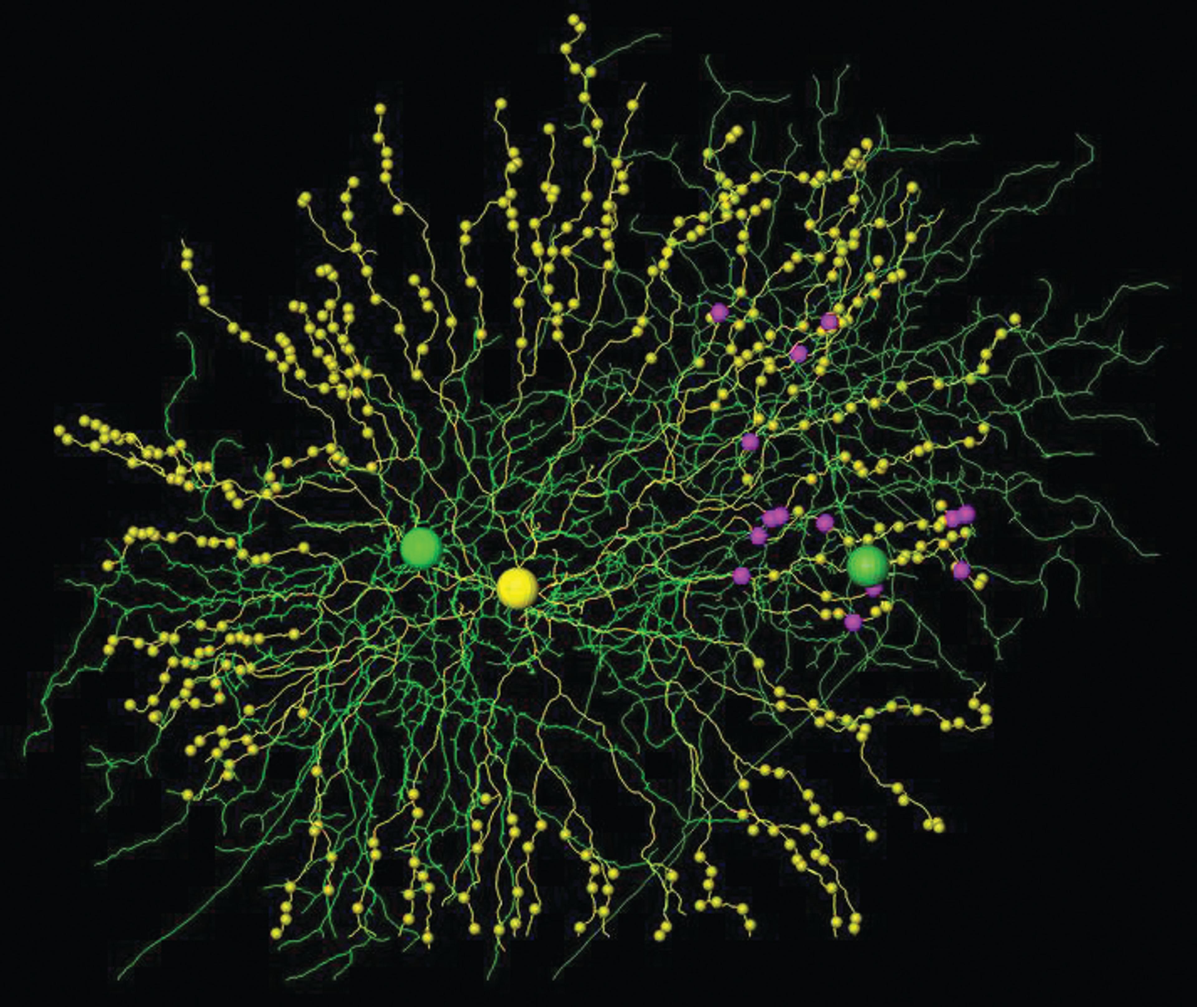
Cells and synapses reconstructed from serial block face electron microscopy data. A single starburst amacrine cell (yellow, note synaptic varicosities) and two direction-selective ganglion cells (green). Even though there is substantial dendritic overlap with both cells, all connections (magenta) go to the right ganglion cell. (Photo credit: © Kevin Briggman/Max Planck Institute)
A key event in nerve cells as they become excited is the rise in the intracellular concentration of calcium. This can be viewed with calcium-sensitive fluorescent dyes. With two-photon fluorescence microscopy Professor Denk could perform two tasks simultaneously: stimulate the photoreceptors of an isolated rabbit retina by moving a light stimulus over it in one direction, and at the same time, record the fluorescence from the individual dendrites of starburst amacrine cells. Professor Denk found that the starburst amacrine cells generated calcium signals only along those dendrites that were aligned in the same direction as the movement of light. Each dendrite, it seemed, behaves as a separate signalling unit.
The next question was how starburst amacrine cells then convey directional information to certain retinal ganglion cells and not others. One clue came from dual electrical recordings showing that amacrine cells were capable of delivering an inhibitory signal to the "null direction" side of a retinal ganglion cell by releasing the neurotransmitter GABA. And yet the dendrites of each starburst amacrine cell appear – by conventional light microscopy – to reach all around adjacent retinal ganglion cells. Professor Denk reasoned that any asymmetry in the signals from starburst amacrine cells must therefore be due to an as yet undetected imbalance, either in the arrangement of the tiny connections, synapses, formed between the two cell types, or in the types of signals that they deliver – some synapses delivering inhibitory signals more strongly than others.
This was where Denk’s second invention was critical. The electron microscope is an extremely powerful tool for viewing the individual synapses of neurons, at orders of magnitude higher resolution than light microscopy. The high resolution, however, means that only a few synapses on one part of a cell can be studied at any one time, making it difficult to survey all those synapses scattered on widely spaced dendrites that run in different directions. Traditional electron microscopy (EM) involves cutting extremely thin slices of a block of tissue, and viewing individual slices to see parts of a cell that lie at different depths. The individual sections have to be aligned correctly in order to see how the tissue components connect.
Professor Denk’s solution – first published in 2004 - was to perform scanning EM not on the cut sections of a tissue sample, but rather on the surface of the tissue block after repeatedly removing each top section – which he called "serial block-face electron microscopy." This enabled researchers to generate serial images that went deeper into the tissue as each top slice was removed – to follow fine neural processes for hundreds of micrometers. In 2011, Professor Denk reported his breakthrough: there was indeed asymmetry in the numbers, or density, of synaptic connections made between pairs of starburst amacrine cells and retinal ganglion cells that were responding to a directional light signal. It was only at the ultrastructural level of synapses that this asymmetry was apparent – with the synapses from starburst amacrine cells gathered preferentially on the "null side" of the responding retinal ganglion cell. According to Professor Denk, these then deliver a negative signal if light moves in the null direction, and permit the retinal ganglion cell to fire only when the light is moving in the opposite direction.
Professor Denk concluded, “Our study demonstrates how otherwise intractable neurobiological questions can be addressed by combining functional imaging with the analysis of neuronal connectivity using large-scale electron microscopy.”
Ann M. Graybiel, who is Investigator at the McGovern Institute for Brain Research, MIT, has devoted her career to probing the question of how we learn to perform tasks that become repetitive to the point where the actions become almost automatic – such as driving a car and applying the brakes when we see a red light.
But, rather than studying the entire nervous system of a single model organism, Professor Graybiel has studied one brain region – a portion of the basal ganglia known as the striatum – in mice, rats, cats, monkeys, and humans using a whole range of technologies from microscopy through electrophysiological recordings to genetics. The striatum is a globular region (in human beings about an inch wide) that lies deep in the brain and was relatively neglected before Professor Graybiel began her studies. She was intrigued by observations that this region had an abnormal appearance in post-mortem examination of patients with Parkinson’s Disease and Huntingdon’s Disease. It appeared, therefore, to be important to the control of movement – which is typically abnormal in such conditions - but no one knew at this time that the striatum was important for learning.
In the late 1970s, she revealed that the striatum, rather than being an amorphous mass of neurons, instead consisted of discrete clusters of neurons, “striosomes” (surrounded by “matrix”), that were visible upon staining for different neurotransmitters and their receptors. In samples from the brains of humans, cats, and monkeys, she saw patches containing high concentrations of opioid receptor-bearing neurons, surrounded by a dense matrix of nerve fibers rich in acetyl cholinesterase.
Suspecting that the clustering of nerves in the striatum may somehow relate to their function, Professor Graybiel later showed (in monkeys) that neurons in the sensory cortex, where sensation is perceived, project axons into the striatum. Here they cluster together with neurons extending from parts of the motor cortex that trigger movement in the same body parts. Thus, axons conveying sensory information about pain or heat in the finger, for example, project to the same areas in the matrix of the striatum as do axons that trigger movement of the same finger. Professor Graybiel discovered that there are multiple such “matrisomes” for any one body part, in a form of repetition and overlap that others have likened to a “mosaic of broken mirrors”, to form multiple loops connecting the outer layer of the brain – the neocortex – where cognition, perception, and motor control reside, as well as the brainstem, which coordinates movement.

The striatum (red) plays an important role in the learning of new tasks by a ‘use it or lose it’ process of reinforcement of nerve signalling pathways. Abnormalities in the same neural circuits are linked to disorders such as Huntington’s disease, Parkinson’s disease, obsessive-compulsive disorder, and autism. (Photo credit: Anatomography, maintained by Life Science Databases(LSDB), under Creative Commons Attribute.)
Professor Graybiel now had a map – like an electrical wiring diagram – of the routes and connections taken by neurons through the striatum. Her next series of studies was to show that these multiple loops and bundles of neurons form the anatomical basis for the learning of new habits in response to environmental cues. A key experiment was to implant tiny electrodes into the striatum of rats – and monkeys – as they learned new tasks. Rats were trained to search for food in a T-shaped maze. They would have to learn whether turning left or right at T-junctions was going to lead to the reward – Belgian chocolate being the favorite. When the animals first encountered a new maze, a high level of firing was detected throughout the task. But once the animals had learned the best route, the firing pattern was starkly different: high-level electrical activity appeared as the animals entered the maze, and when they ate their reward, but in between – while following the now-learned route in a more automatic fashion – neuronal activity was much lower. Professor Graybiel termed this neuronal activity marking the ends of a sequence of actions “chunking,” and suggested that this reflects the mastering of a learned skill. She made similar findings in monkeys, who learned to make a specific set of eye movements, and other behavioural responses in return for a reward such as a drink of juice. The pattern of bursts in neuronal firing, at the beginning and end of a learned task, marks the start and finish of a behavioral unit – in other words, the set of the actions needed to perform a task.
In 2009 and 2010, Professor Graybiel and her team reported follow up studies in mice and rats being trained to find food in a T-maze. They detected the development of different firing patterns in different parts of the striatum, and between individual neurons and clusters (ensembles) of neurons. The studies support the notion that the striatum is not only important for establishing the sequence of actions involved in a particular behavior, but also that it is flexible enough to allow additional influences – such as thoughts and emotions. In other words, the striatum is key to both making and breaking habits.
Professor Graybiel’s work also supports the idea that learning new tasks involves a “use it or lose it” process of selecting and reinforcing particular nerve signalling pathways at the expense of others, in order to arrive at the most rewarding behavior. In a 2011 interview, Professor Graybiel commented on actions such as riding a bike and playing tennis: “These patterns and many others in our behavior probably are developed partly through instruction and partly through trial and error learning….these basal ganglia are par excellence trial and error learning devices.” She suspects, too, that the same neural circuits may be going into “overdrive” in producing the repetitive behaviors found in disorders such as obsessive-compulsive disorder, Tourette’s Syndrome, autism, and schizophrenia.
The importance of the striatum in learning appears to be conserved throughout evolution – the equivalent region in birds is important to bird song. In humans, the use of PET scanning and functional magnetic resonance imaging (fMRI) has revealed abnormalities in the striatum in patients with obsessive-compulsive disorder and Tourette’s Syndrome.
Some of Professor Graybiel’s more recent work has centered on findings that certain intracellular signalling molecules are more highly expressed in parts of the striatum of mice and rats during the learning of new habits. She is also exploring their role in animal models for human conditions including Huntington’s disease, Parkinson’s disease, and drug addiction.