


2014 kavli prize in Neuroscience
2014 Kavli
Prize in
Neuroscience

The Norwegian Academy of Science and Letters has decided to award the 2014 Kavli Prize in Neuroscience to Brenda Milner, John O'Keefe and Marcus E. Raichle.
“For the discovery of specialized brain networks for memory and cognition."
Committee Members
- Ole Petter Ottersen (Chair), Centre for Molecular Biology and Neuroscience, University of Oslo, Norway
- Cornelia Bargmann, The Rockefeller University, USA
- Lily Jan, University of California, San Francisco, USA
- Erwin Neher, Max Planck Institute for Biophysical Chemistry, Germany
- Stanislas Dehaene, INSERM-CEA, France
Citation from the Committee
The higher cognitive functions of our brains such as attention, memory, and planning are essential to our ability to create rich mental lives. The three Kavli laureates discovered that these functions are produced by specialized brain systems, which they analyzed at different levels – from single neurons to brain regions and interconnected networks.
Brenda Milner discovered regions of the brain specialized for memory formation and other cognitive functions. She found that HM, a neurological patient with damage to the hippocampus and surrounding regions, could not acquire new memories of events, but could speak, reason, and recall long-past memories. By studying this patient and others, she discovered that the medial temporal lobes are needed to form one kind of memory, which we now call episodic memory, and not for other kinds of memory like procedural memory. She made similar discoveries of specialized functions within the frontal lobes for planning and organizing behavioral sequences.
John O’Keefe discovered that the hippocampus contains neurons that encode an animal’s specific location. These place cells allow detection of novelty and changes in familiar environments and collectively form a cognitive map critical for animal navigation behaviour.
This discovery provides a sterling example of neuronal signalling in a specific brain region involved in memory formation.
Marcus E. Raichle designed methods for visualizing the activity of the normal living human brain. These techniques permitted the quantitative measurements of blood flow and metabolism in localized regions of the brain and provided the basis for all modern functional imaging studies. They allowed mental operations such as reading, attention, and memory to be associated with activity in specialized networks of brain regions, present in all human brains. Raichle’s observation of systematic patterns of ongoing brain activity when the subject is in a resting state has transformed the way the human brain is now being studied in health and disease.
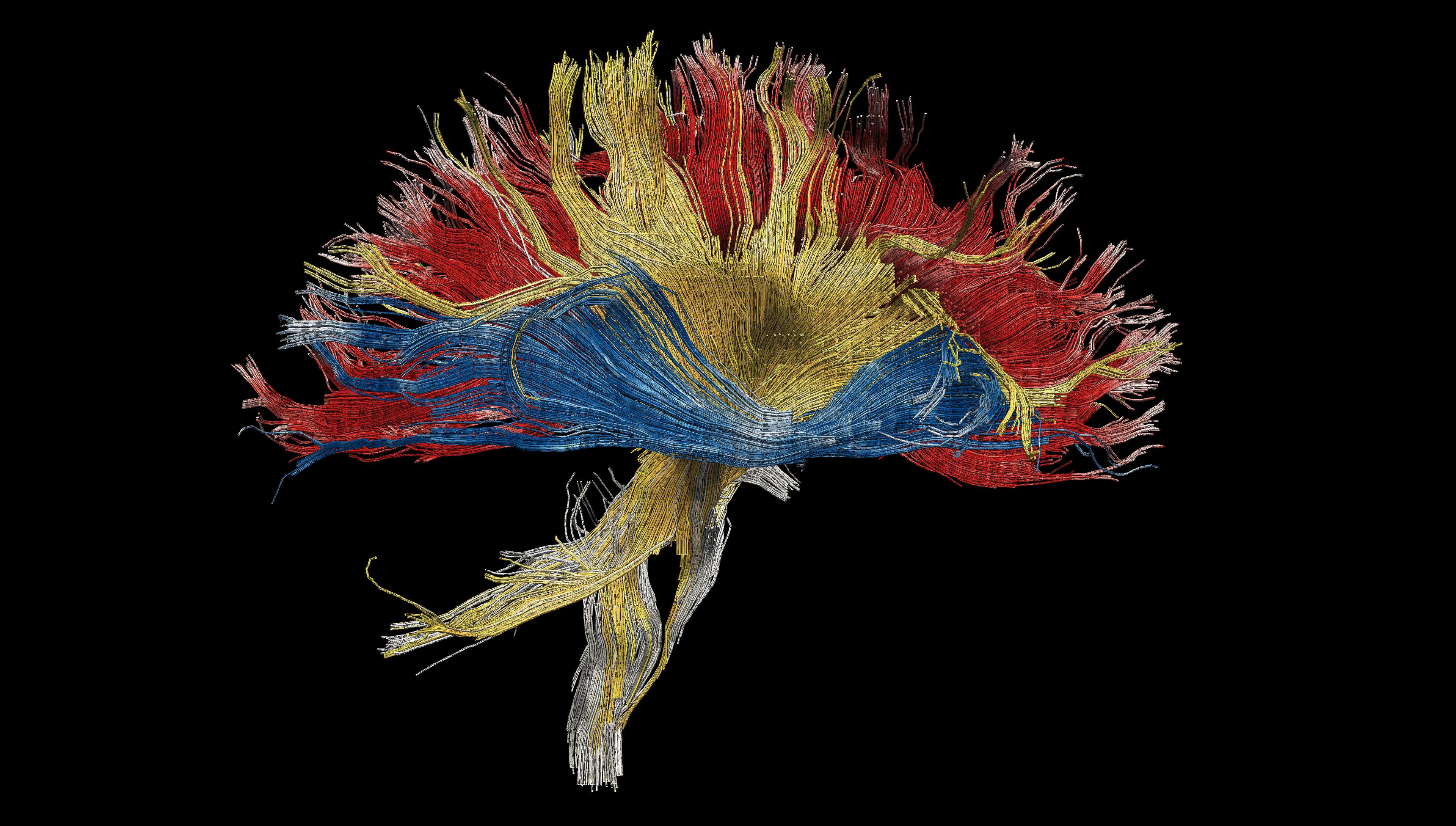
A reconstruction of pyramidal neurons (Photo credit: © Professor Michael Hausse).
The 2014 Neuroscience Kavli Prize explained
The functions of our brains such as attention, memory, and planning are important in our ability to create rich mental lives. Memory is an essential component of being human, from the recognition of where we are, through learning new skills, to being able to recall events. In humans, memory can be said to define who we are; we know that loss of memory can have devastating effects on a person’s personality. One of the great challenges for researchers studying the human brain is working out which areas of the brain are involved in specific activities, and this in itself poses a further challenge: how to measure activity in the brain whilst humans are behaving normally in a way that can be repeated and compared.
By Matt Wakelin
The recipients of the 2014 Kavli Prize in Neuroscience have played major roles in advancing our understanding of memory and in the development of techniques to measure brain activity. They have discovered that these functions are produced by specialised brain systems, which they have analyzed at different levels and using different approaches. They have made significant contributions to our understanding of the cells and regions of the brain involved in memory and cognition.

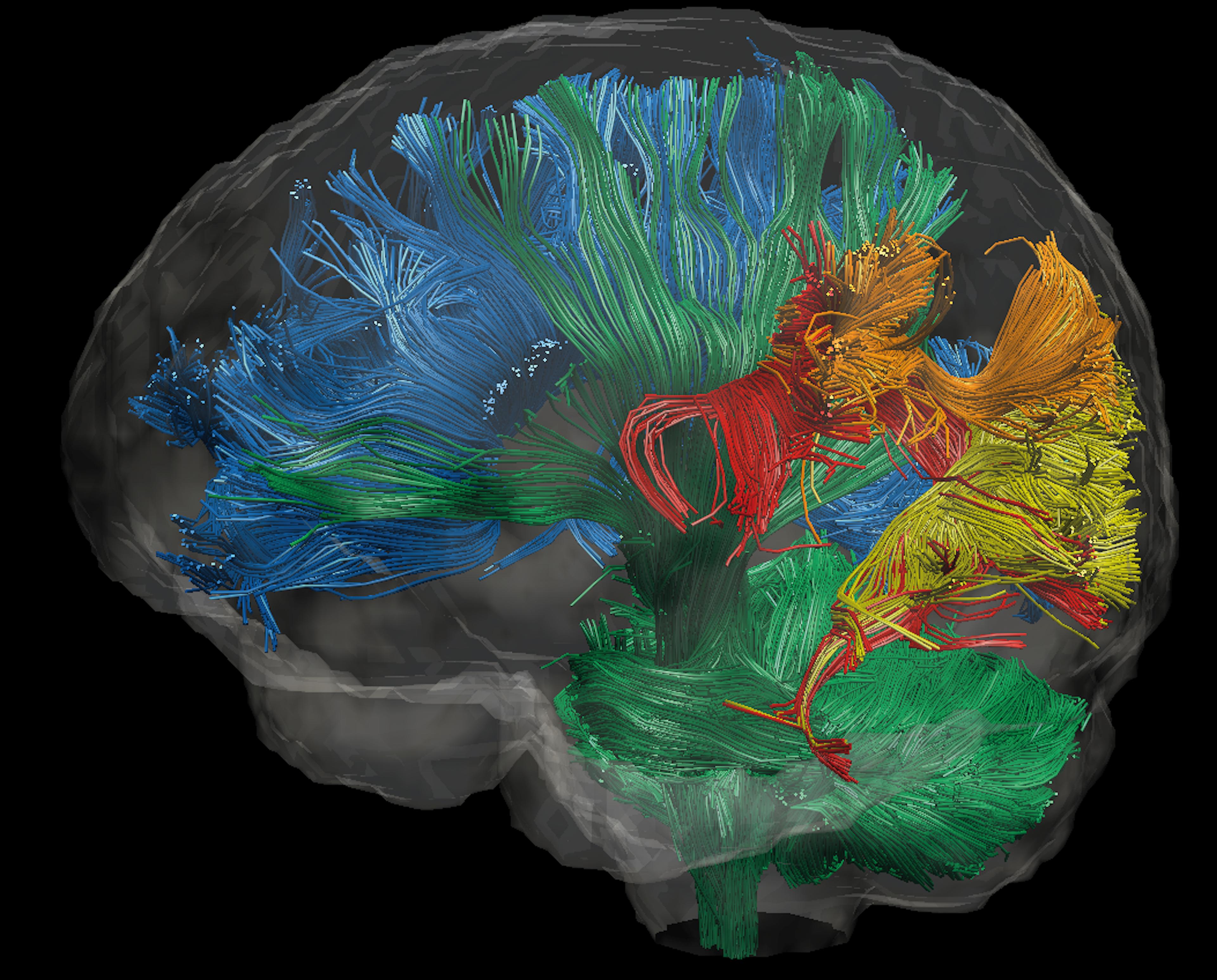
The brain is made up of different regions, each with specialised functions: White matter tracts throughout the human brain (Photo credit: © Dr Jamie Kawadler).
Brenda Milner, a neuropsychologist at the Montreal Neurological Institute, McGill University, is a pioneer in the field of neuropsychology and in the study of memory and other cognitive functions in humankind. She discovered regions of the brain specialized for memory formation and other cognitive functions. Much of her work has been built on observation of patients with memory deficits, and as she describes herself, her success is due partly to being what she calls "a noticer." “The thing that has driven me my whole life is curiosity. I am incredibly curious about the little things I see around me”. Milner pioneered an entirely new scientific discipline – which Nobel Prize winner Eric Kandel described as creating the new field of cognitive neuroscience by merging neurology and psychology. Her detailed and long-term studies of patients, lasting for many years, before and after brain surgery, have made significant contributions to the understanding of the structure of the brain, especially the functions of the hippocampus and the temporal, frontal and parietal lobes in learning, memory, and speech functions.
Brenda Milner studied the effects of brain damage, in particular on a region known as the medial temporal lobe, and its effects on memory. Her most famous work was based on studies of Henry Molaison, formerly known as patient HM. Henry Molaison suffered from epilepsy that has sometimes been attributed to a bicycle accident at the age of seven. In 1953, he underwent a major operation to remove parts of the brain as treatment for his epilepsy, including large portions of the hippocampus and adjacent structures. Although the surgery was deemed a success in controlling the seizures associated with his epilepsy, Henry Molaison was found to suffer from amnesia; he had lost the ability to convert short-term memory into long-term memory.
Through rigorous experiments, Milner discovered that Henry Molaison could learn and remember particular types of tasks, for example new motor skills, although he could not remember learning the skill, and that his memories of the past before the operation were seemingly intact. In the early stages of her work with him, Milner wanted to completely understand his memory impairments. She showed that the medial temporal lobe amnestic syndrome is characterized by an inability to acquire new memories and an inability to recall established memories from a few years immediately before damage, while memories from the more remote past and other cognitive abilities, including language, perception, and reasoning were intact. For example, Milner spent three days with HM as he learned a new perceptual-motor task in order to determine what type of learning and memory were intact in him. This task involved reproducing a drawing of a star by looking at it in a mirror. His performance improved over those three days. However, he retained absolutely no memory of any events that took place during those three days. This led Milner to speculate that there are different types of learning and memory, each dependent on a separate system of the brain. She was able to demonstrate two different memory systems – episodic memory and procedural memory.
Milner’s research on HM, working with William Scoville, was first reported in 1957, in the Journal of Neurology, Neurosurgery and Psychiatry, which presented results “…which point to the importance of the hippocampal complex for normal memory function.”
Near the end of his life, Molaison regularly completed crossword puzzles – provided that the clues referred to pre-1953 answers. Through her encounters with him, Milner established that people have multiple memory systems, governing different activities like language or motor skills, opening the way for a greater understanding of how the brain works. Her work has made a major contribution to our understanding of the role that particular regions of the brain play in processing our memories and organising information. Furthermore, her research has also influenced the development of tests to assess, diagnose, and treat people with brain disorders resulting from traumatic injury and degenerative diseases, as well as from psychiatric illness.
Milner stated in an interview with the McGill Journal of Medicine: “To see that HM had learned the task perfectly but with absolutely no awareness that he had done it before was an amazing dissociation. If you want to know what was an exciting moment of my life that was one”. Although they worked together for more than three decades, HM was never able to remember Brenda’s name.

A reconstruction of pyramidal neurons (Photo credit: © Professor Michael Hausse).
John O’Keefe is a professor of cognitive neuroscience in the Department of Cell and Developmental Biology at University College London. Movement is integral to human existence; as well as being able to physically move from location to another, we also have the mental capacity to imagine where we are.
John O’Keefe has illuminated key aspects of these remarkable navigational and conceptualizing abilities. Navigation is a complex activity. It requires integration of visual information, as well as memory and planning. John O’Keefe discovered that certain cells in a region of the brain called the hippocampus preferentially fired, or were activated, when an animal was in a particular environmental location – the first description of "place cells." This led Professor O’Keefe to make the initially controversial but ultimately influential suggestion that the hippocampus held some kind of "cognitive map" of the outside world.
The hippocampus, from the Greek hippos meaning “horse,” and kampos meaning “sea monster,” is a region of the brain so named due to its resemblance to a seahorse. Although very small (less than the size of a little finger), it plays a vital role in brain function. The hippocampus is responsible for both our short-term and long-term memory, and also for spatial navigation. Despite its small size, the hippocampus is complex; it is made up of about 40 million nerve cells, with each one of these cells able to connect with up to 10,000 other cells. It can be likened to a complicated circuit board that sends information to other parts of the brain. We know that the hippocampus is one of the first regions of the brain to suffer damage in neurodegenerative disease, resulting in memory loss and disorientation.
John O’Keefe, along with John Dostrovsky, discovered in 1971 that the hippocampus contains special nerve cells that are involved in determining an animal’s specific location, which they called place cells. This ground-breaking work depended on O’Keefe and Dostrovsky being able to record the firing of a single neuron in the hippocampus during normal behavior. Small electrodes were implanted in a rat’s brain; after recovery from surgery, the electrodes were monitored. When an electrode is close to a neuron, it can record the current produced by the neuron when it fires. With a computer data-acquisition system, the timing of a neuron’s firing pattern, combined with information about the location of the animal, can be captured. In this way, a firing-rate map could be generated, showing the position of an animal when the cell fires. Imagine hundreds of place cells, covering all locations of accessible space. This concept led O’Keefe to propose that the hippocampus acted as a "Cognitive Map" that represented environmental space and guided efficient navigation in rats and, presumably, humans.
A cognitive map is a device for representing an animal’s current environment, its position within it, and the location of both desirable objects, such as food and threats that should be avoided. This cognitive map can direct the animal’s behaviour on the basis of distances and directions towards desired goals or away from undesirable objects and the locations. In addition, the cognitive mapping system detects the absence of representations of novel environments and changes in maps of familiar environments and uses these mismatches to trigger and control exploration.
In 1978, working with Lynn Nadel, O’Keefe expanded this notion and suggested that in addition to the place cells, the hippocampus might also contain information about direction and distance. The hippocampus can receive information from many different sensory inputs to help build the cognitive map. This work showed that place cells allow the detection of new environments and also changes in familiar environments, in essence forming a map of the surrounding environment within an animal’s brain, termed a cognitive map.
John O’Keefe has continued his work on how the cognitive map theory can be expanded to explain the episodic memory deficit in patients with hippocampal damage. Working with Neil Burgess, he has used computational models of the cognitive map to generate theoretical predictions of hippocampal function. These theoretical predictions can then be studied experimentally.
As he noted in an interview in 2013, the hippocampus may play a role in episodic memory. Episodic memory involves the ability to learn, store, and retrieve information about unique personal experiences that occur in daily life. These memories typically include information about the time and place of an event, as well as detailed information about the event itself. The ability to describe the details of your first day at school, a recent holiday gathering, or office meeting that took place in the previous weeks or months, for example, depends heavily on intact episodic memory function. It is known that one of the first noticeable symptoms displayed by Alzheimer’s patients is a deficit in their navigational ability; they tend to get lost in familiar environments. Knowing how this memory function should work in healthy people opens the door to understanding what has changed in patients with memory loss.
Marcus E. Raichle, a neurologist, is a Professor of Radiology, Neurology, Neurobiology, and Biomedical Engineering at Washington University in St Louis. Through his work with his colleagues, he has made outstanding contributions to the study of the human mind through the development and use of two techniques in the study of the brain: positron emission tomography (PET) and functional magnetic resonance imaging (fMRI).
PET works by measuring the signals given off by a radioactive material, or radioisotope, that is injected into the body. The radioisotope can be used to show metabolic activity in different tissues. Using PET, a three-dimensional image of functional processes in the body can be developed.
fMRI is a technique for measuring brain activity. It works by detecting the changes in blood oxygenation and flow that occur in response to neural activity – when a brain area is more active it consumes more oxygen, and to meet this increased demand blood flow increases to the active area. fMRI can be used to produce activation maps showing which parts of the brain are involved in a particular mental process.
The great advantage of these two techniques is that they allow study of the normal living human brain in action. By allowing quantitative measurements of blood flow and metabolism in regions of the brain, measurements that can be determined objectively, and they allow the complex behavioural patterns of humans to be broken down into their component processes. These techniques have allowed scientists to investigate mental operations in humans, such as reading or memory, and to link these to activity in particular regions of the brain. In this area, Marcus Raichle’s contribution has been preeminent.
Raichle first used radioisotope techniques to study the human brain during his neurology residency at New York Hospital – Cornell Medical Center. He continued these studies when he joined Washington University School of Medicine. As he stated himself, he was interested in using tracers in the brain as they provided the possibility of taking objective measurements in real time. However, these early studies were invasive and limited the types of studies that could be carried out. He looked to other, less invasive approaches that could be used and soon focussed on PET, which allows researchers to safely and non-invasively study the living human brain and track and record its function in health and disease.
His landmark study in 1988 described the first integrated strategy for the design, execution, and interpretation of functional brain images. It represented 17 years of work developing the components of this strategy (e.g. rapid, repeat measurements of blood flow with PET; stereotaxic localisation; imaging averaging; and a cognitive subtraction strategy). Another seminal study led to the discovery that blood flow and glucose utilisation change more than oxygen consumption in the active brain (Science, 1988) causing tissue oxygen to vary with brain activity. This discovery provided the physiological basis for subsequent development of fMRI and caused researchers to reconsider the dogma that brain uses oxidative phosphorylation exclusively to fuel its functional activities.

Imaging can reveal the flow of information in the brain: MRI-based imaging of six of the major white matter pathways in the human brain (Photo credit: © Dr Jamie Kawadler).
In addition to pioneering these techniques, Raichle has also been instrumental in developing computer programs to make the most of PET imaging and be able to study regions of the brain to an accuracy of one millimeter.
Raichle’s most recent research has helped in the development of a much better understanding of those areas of the normal human brain responsible for language, thought processing, and emotion. By using PET to monitor blood flow and metabolism, Raichle and his collaborators have shown how the brain responds when a subject is asked to perform tasks as diverse as memorizing words or anticipating an unpleasant experience. In addition, they have mapped areas involved in attention, analyzed chemical receptors in the brain, and investigated the physiology of major depression.
Raichle’s observation of patterns of ongoing brain activity when the subject is in a resting state, or when the brain is not actively engaged in performing tasks such as recalling events or learning new words, has transformed the way the human brain is now being studied in health and disease. Previously scientists had focused on studying the active brain, but Raichle’s work opened up the possibility that studying the resting brain might help in the understanding of how the brain works. Seeking to explain why tasks might decrease activity in the brain, they employed an innovative strategy to define a physiological baseline (PNAS, 2001). This work had profound implications for using imaging and the understanding of how the brain processes information. This work led to the concept of a "default" mode of brain function and invigorated studies of intrinsic functional activity, an issue largely dormant for more than a century. An important aspect of this work was the discovery of a unique neural network in the brain that has come to be known as the default network – a network of parts of the brain that are active when someone is involved in internal thoughts, such as daydreaming or retrieving memories. This network is now the focus of work on brain function in health and disease worldwide, and it has been hypothesized to be relevant to disorders including Alzheimer’s disease, autism, and schizophrenia.