


2016 kavli prize in Neuroscience
2016 Kavli
Prize in
Neuroscience
The Norwegian Academy of Science and Letters has decided to award the 2016 Kavli Prize in Neuroscience to Eve Marder, Michael M. Merzenich and Carla J. Shatz.

“For the discovery of mechanisms that allow experience and neural activity to remodel brain function.”
Committee Members
- Ole Petter Ottersen (Chair), Centre for Molecular Biology and Neuroscience, University of Oslo, Norway
- Cornelia Bargmann, The Rockefeller University, USA
- Erwin Neher, Max Planck Institute for Biophysical Chemistry, Germany
- Susan McConnell, Stanford University, USA
- Antoine Triller, Institut de Biologie de l'École Normale Supérieure, France
Citation from the Committee
How does the brain change during learning and development, while remaining structurally stable and producing reliable behaviour? This fundamental question has been addressed by the three 2016 Kavli Prize laureates in Neuroscience.
Their discoveries showed how neuronal activity, generated either by experience or by intrinsic brain function, actively sculpts structural and functional connections between nerve cells. At the same time, essential stability is provided by self-regulating mechanisms that drive nerve cells to produce consistent patterns of activity.
Michael Merzenich demonstrated that sensory circuits in the cerebral cortex can be reorganized by experience in adulthood. Different parts of the body are represented in a continuous map in the somatosensory cortex. After demonstrating reorganization of this map after injury, Merzenich showed that simply expanding or limiting the use of different fingers leads to a corresponding change in the representation of the hand in the brain. Similarly, he showed that the auditory cortex can change its map of sound frequencies after individuals are trained to detect fine differences in pitch. This discovery helps explain how humans can recover their perception of speech with electronic cochlear implants, which generate signals much simpler than normal auditory inputs. Merzenich showed that neuromodulators as well as cognitive factors including attention determine whether adult plasticity takes place. This work is being extended in humans to maximize learning and recovery from brain injury and disease.
Carla Shatz showed how patterns of activity in the developing brain instruct and refine the arrangement of synapses between neurons. She demonstrated that the formation of appropriate connections between the eye and the brain of mammals depends on neuronal activity before birth. She discovered that spontaneous waves of activity sweep across the retina early in development, and showed that these organized activity patterns select the final set of connections from a coarse, genetically-determined map. Her demonstration that “neurons that fire together, wire together” links the mechanisms of brain wiring during development to those underlying adult learning and memory.
Eve Marder used the simple circuits of crustaceans to elucidate the dynamic interplay between flexibility and stability in the nervous system. She showed that numerous neuromodulators reconfigure the output of adult neural circuits without altering their underlying anatomy. At the same time, she found that circuits can generate similar neuronal and network outputs from many different configurations of intrinsic neuronal excitability and synaptic strength. This apparent paradox was solved by her recognition that neurons have a self-regulating homeostatic programme that drives them to a stable target activity level. With the other two Kavli Prize laureates, Marder defined the mechanisms by which brains remain stable while allowing for change during development and learning.
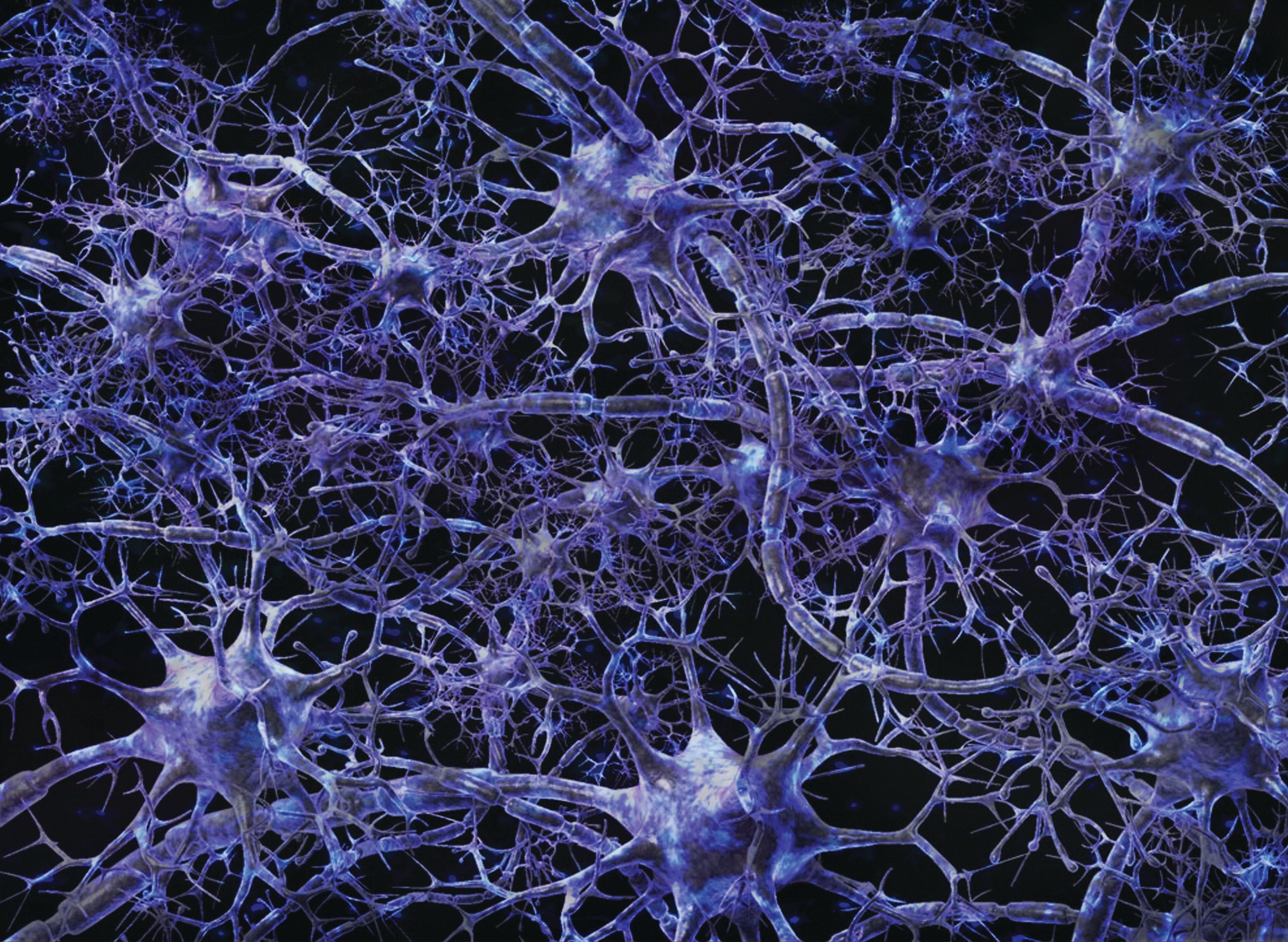
Neural network. Dense network of myelinated nerve cells. (Credit: Arran Lewis, Wellcome Images)
The 2018 Neuroscience Kavli Prize explained
Early 20th century pioneers of neuroscience used the power of microscopy to marvel at the intricate wiring of the brain and peripheral nerves. They photographed and traced the long processes and connections and saw that these took shape early in life and then apparently remained fixed. The brain, it seemed, was hardwired and inflexible once we reached adulthood. This appeared to match the notion that it is easier for children to learn new skills such as a language or musical instrument than it is for adults. Adults seem to have to work harder at learning new skills and memorising information, but no one understood why.
By Julie Clayton
Over the past 40 years, however, the three Kavli neuroscience prize-winners of 2016 have challenged these assumptions and provided a convincing view of a far more flexible adult brain than previously thought possible – one that is ‘plastic’, or capable of remodelling. Working in different model systems, each researcher has focused on how experience can alter both the architecture and functioning of nerve circuits throughout life, given the right stimulus and context. They have provided a physical and biochemical understanding of the idea of ‘use it, or lose it’. This new picture of a more adaptable brain offers hope for developing new ways to treat neurological conditions that were once considered untreatable.
Embarking on his research career in the late 1970s, Michael Merzenich was interested in how the brain’s outer layer, the cerebral cortex, located and responded to sensory information. Using microelectrodes to monitor individual neurons in the brains of owl monkeys, he found that the somatosensory cortex has a complete topographical ‘map’ of the entire body surface, in which adjacent areas of the map responded to body parts that were next to one another, such as the fingers. Each point on the map was a cluster of neurons responding to stimulation of a different patch of skin.

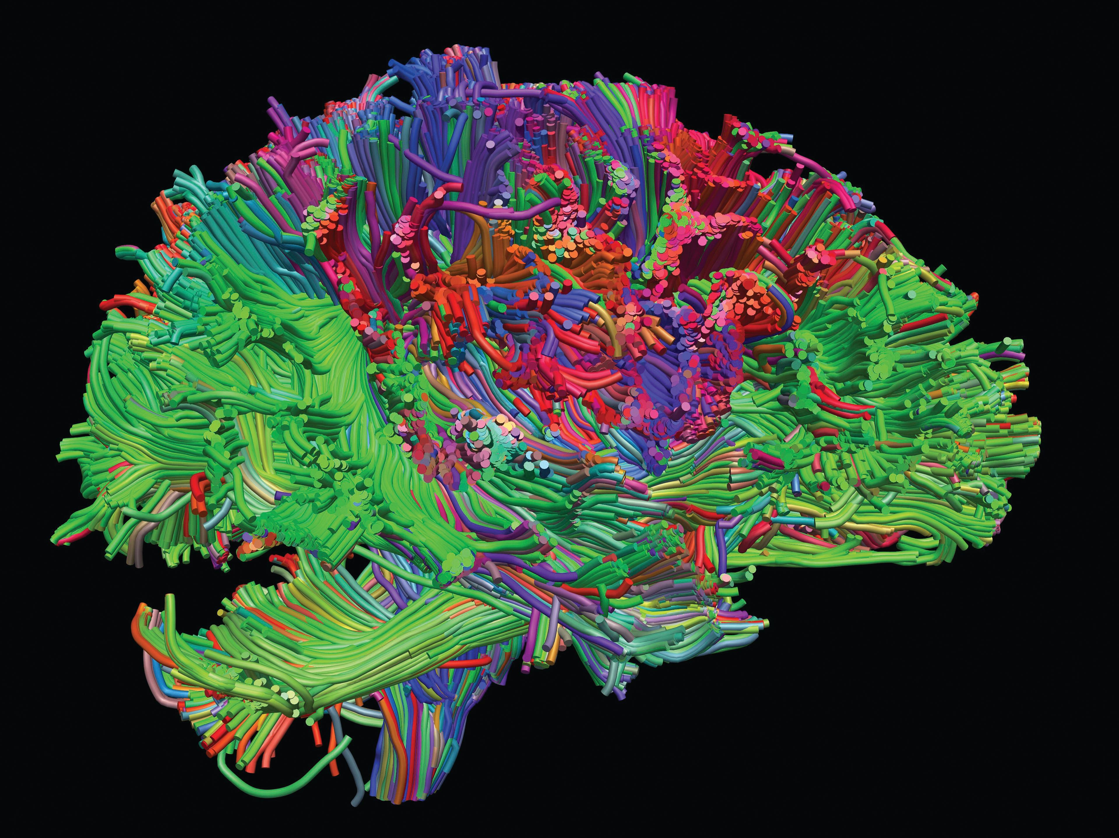
Healthy adult human brain viewed from the side. The brain is viewed as if looking through the head from a person’s right ear. Brain cells communicate with each other through these nerve fibers, which have been visualised using diffusion imaging tractography. (Credit: Henrietta Howells, NatBrainLab, Wellcome Images)
Previous work by others had shown that nerve damage, or limb amputation, led to a reorganisation of neuronal pathways in the cortex of very young animals. Merzenich questioned whether the same could be true for adults, and during the next two decades performed studies which revolutionised understanding of the mature brain. Studying owl monkeys and squirrel monkeys, he found that damage to a nerve, or loss of one or two fingers caused the portion of the map that had lost its input to be overtaken by neighbouring maps, as neurons in the affected area became responsive to other areas of skin. It was rather like redrawing the political boundaries on a map of the world after a power struggle – borders shifting as some territories expanded while others shrank.
Conversely, when the monkeys had two fingers strapped together for some months, the cortical maps for the two fingers merged, as though they were for one finger. But after separating the two fingers again, their individual representations reappeared.
Provocatively, Merzenich concluded that the potential for brain remodelling, or plasticity, was not lost beyond childhood, but could be switched on again in adulthood. It was a physical explanation for something that psychologists had known for decades – that the adult brain is capable of learning new tasks and compensating for brain damage, with the right training and therapy.
Merzenich extended his findings to sound perception, showing that the auditory cortex also forms sensory maps corresponding to different sound frequencies, and that these maps are equally malleable in adulthood. For example, in monkeys that learnt to associate certain sound frequencies with a food reward, the auditory maps representing those frequencies became enlarged in trained animals compared to untrained controls.
These findings helped lead to the development of prototypes of the electronic cochlea implants now available today. These stimulate areas of the brain that would normally respond to the different sound frequencies of human speech.
In another twist, Merzenich and co-workers found that they could trigger the rearrangement of the auditory maps by exposure to a combination of sound frequencies plus electrical stimulation of the part of the hindbrain called the nucleus basalis. This is involved in learning and memory, especially when attention is heightened through fear, or anticipation of reward, and involves the release of the neurotransmitter acetylcholine.
Merzenich believes that the machinery and conditions required for brain plasticity are permanently switched on during childhood, and can be switched on again in adulthood. The necessary circumstances, he suggests, include ensuring that a person’s attention is focused on a task, and that they are experiencing either motivation or success. Merzenich is now applying his basic research findings to the development and testing of new tools for improving brain function in people with a range of neurological and psychiatric conditions, such as Alzheimer’s disease or schizophrenia, or following brain injury.
Carla Shatz has elegantly revealed how the brain is sculpted in early life, both before and after birth, as animals make the transition from the protective environment of the womb to the vibrant and stimulating outside world.
In the late 1970s, Shatz followed up on the Nobel Prize-winning work of Hubel and Wiesel showing that soon after birth, in monkeys and cats, light stimulation of the eye promotes the self-organisation of the visual cortex – the part of the brain responsible for vision. Neurons projecting from the retina cluster into a series of bands across the visual cortex; each band is either left- or right-eye responsive. Closure of one eye during a critical ‘sensitive period’ of several days after birth disrupts this pattern as the bands corresponding to the deprived eye shrink in relation to those receiving signals from the active eye.
Little was known, however, about what happened before birth, when the eyes have yet to be fully developed. Shatz focused on the lateral geniculate nucleus (LGN), where nerves projecting from the eye first become sorted into an orderly set of layers. Shatz’s first major scientific finding was that this layering of neurons in the LGN began before birth, in response to repeated bursts of spontaneous firing by retinal ganglion cells, which spread in waves across the retina. The size of the LGN layers, and the connectivity between neurons within the layers, depended on the intensity of the spontaneous electrical activity.
Shatz wanted to understand in more detail what stabilised these arrangements of neurons in the visual cortex as the brain matures. In 2000, her team found the factor BDNF to have a role. Animals reared in the dark in the first few days of life had reduced levels of expression of BDNF in the visual cortex. Blocking BDNF activity during this time reduced the fine sculpting of layers in the LGN, and bands in the higher visual cortex. Because the release of BDNF from nerve endings is known to strengthen synapses (the connections between neurons), this could provide the ‘on switch’ that promotes the remodelling of neural pathways.

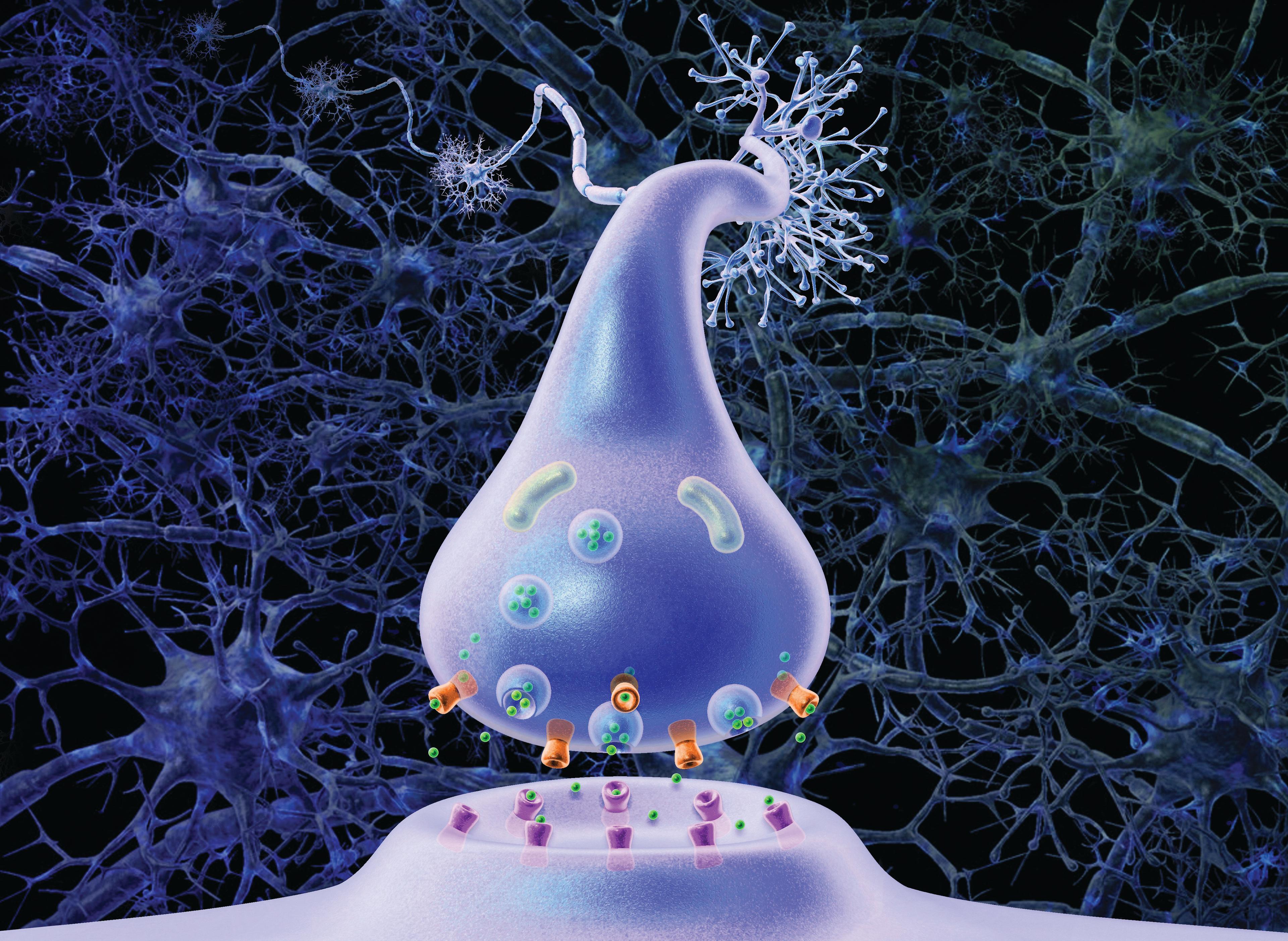
Illustration showing the action of neurotransmitters such as serotonin and noradrenaline in the synaptic cleft. Vesicles containing the neurotransmitter (green) move towards the pre-synaptic membrane where they fuse with the cell membrane, releasing their contents into the synaptic cleft. The neurotransmitter molecules act on the post-synaptic cell by binding to specific receptors on the cell surface (purple). They can also be taken back up by the presynaptic cell via other receptors (orange) for re-use. (Credit: Arran Lewis, Wellcome Images)
Shatz set out to investigate which other genes might be involved in controlling brain plasticity after birth. Surprisingly, she found that some of the active genes were ones that were better known for their role in the immune system, where they help fight infection. Known as MHC (Major Histocompatibility Complex) genes, they code for cell surface proteins. Shatz revealed that in mice which were deficient in certain MHC genes, their visual neurons were more randomly arranged and had many more synapses. Their brain remodelling seemed to be constantly switched ‘on’.
Subsequently, Shatz and colleagues identified a receptor on neurons called PirB (paired immunoglobulin-like receptor B), through which MHC molecules normally delivered the ‘off’ signal. Mice genetically engineered to lack PirB, or treated with an infusion of soluble PirB directly into the brain, also showed an increase in synaptic density and signalling in the visual cortex. Curiously, in a mouse model of Alzheimer’s disease, PirB serves as a receptor for the beta-amyloid protein that accumulates during the disease. Thus signalling through PirB could be responsible for the loss of synaptic plasticity and memory in these animals.
These findings have led Shatz and others into a new line of research on the role of MHC molecules in learning and memory, and how they may contribute to conditions such as stroke damage, Alzheimer’s disease, autism and schizophrenia. It may also be that these molecules provide a possible link between these disorders and viral infections in early life.
Eve Marder has made it her life’s work to understand the properties of neural circuits controlling digestion in lobsters and crabs. It may sound like a rather esoteric choice, but to Marder it is a way to work out the more general rules by which the nervous system in different animals produce rhythmic behaviours including breathing, walking and swimming. She has revealed that neural circuits can be both flexible in response to stimuli and stable over time.
Marder’s focus on the movements of the crustacean gut was due to the exceptional ease with which the entire neural circuit - the stomatogastric ganglion (STG) - could be removed intact from the body and kept alive in the laboratory. It has a well-defined anatomy, with about 30 neurons, 24 of which innervate muscles, and six which cross-link to other ganglia. In the laboratory, this circuit continues to show spontaneous bursts of coordinated electrical impulses for many hours. In the animal, it elicits a pacemaker-like rhythm that prompts gut muscles to contract and relax rhythmically.
Crustaceans do not chew their food in the way vertebrates do - instead they tear and swallow large coarse pieces of food which are moved along the oesophagus to the stomach-like cardiac chamber, where calcified teeth-like projections - known as the gastric mill - crush, cut and grind the food. The finer food particles then pass through the pyloric valve into the pyloric chamber and beyond. The STG is part of a wider stomatogastric nervous system that controls the passage of food along the full length of the digestive tract. Between 100 and 250 nerve fibres project onto the STG from the brain and spinal cord, enabling central nervous system (CNS) regulation.
Marder wanted to understand how the STG was regulated by external influences, such as hormones or substances released by CNS nerve fibres projecting onto the STG, or produced by STG neurons. Others had shown that the neurotransmitter glutamate was released by some STG motor neurons. Marder’s PhD work - which earned her a paper in the journal Nature - was to reveal that a second neurotransmitter, acetylcholine, was released by other STG neurons. This finding set the scene for further studies to identify how neurotransmitters and neuromodulators might modify the rhythmic firing of the STG. During the 1980s, Marder led studies using antibodies and the technique of immunocytochemistry to reveal the presence of many different neuromodulators in nerve fibres around the STG of crabs, including serotonin, proctolin and GABA.
Electrophysiological experiments then led to further significant findings including the first demonstration that two different neurons that were electrically coupled could produce different neurotransmitters. The neuromodulator dopamine, for example, affected STG neurons in different ways - inhibiting some and stimulating others. In this way, Marder was able to work out key principles about the regulation of neural networks, including the fact that the same neuromodulator can have differential effects in different parts of the network.
But why were so many neuromodulators involved in regulating such a relatively small number of neurons? Marder hypothesised that this enabled a whole range of effects to be elicited which fine-tune the end behaviour. It meant that that neuronal circuits, once thought to be hard-wired and fixed in their firing patterns, are capable of considerable variation in output. Neuromodulation, wrote Marder, ‘adds extraordinary richness to the dynamics that networks can display.’
Neural network. Dense network of myelinated nerve cells. (Credit: Arran Lewis, Wellcome Images)
Another of Marder’s key contributions was the co-development of the dynamic clamp tool, in 1993. This allows computer-controlled stimuli to be applied to neurons in culture, and measures their effects on cell membrane conductance. This enables the development and testing of computational models of neural circuits. Lately, Marder has scrutinised how neural circuits can tolerate perturbations to individual neurons and remain functionally stable. Her team is currently examining the ion channels that open and close to allow the flow of ions as electrical impulses pass along the nerve cell membrane. Changes in ion channel expression, induced by neuromodulators, can change the polarisation of the cell membrane and its electrical conductance. This, in turn, affects the magnitude of release of neurotransmitter at the nerve terminal and the strength of muscle contraction.
Marder hopes eventually to have a fuller picture of how neural circuits maintain their robustness over time despite the changes induced by neuromodulators and the routine turnover of membrane and other cell components throughout life. Such insights may also contribute to the understanding of functional decline in different neurological conditions.