


Numerous Possibilities
As told by John O'Keefe
I was born in November 1939 in Harlem, New York to Irish immigrant parents and grew up in the South Bronx. My parents had landed in New York on the eve of the depression; my father’s hope was that I would never have to live through one myself. My mother’s passage was funded by an uncle and redeemed by seven years’ indentured labour. Neither had completed elementary school in Ireland, but my father studied in the evening in New York and attained a high school degree.

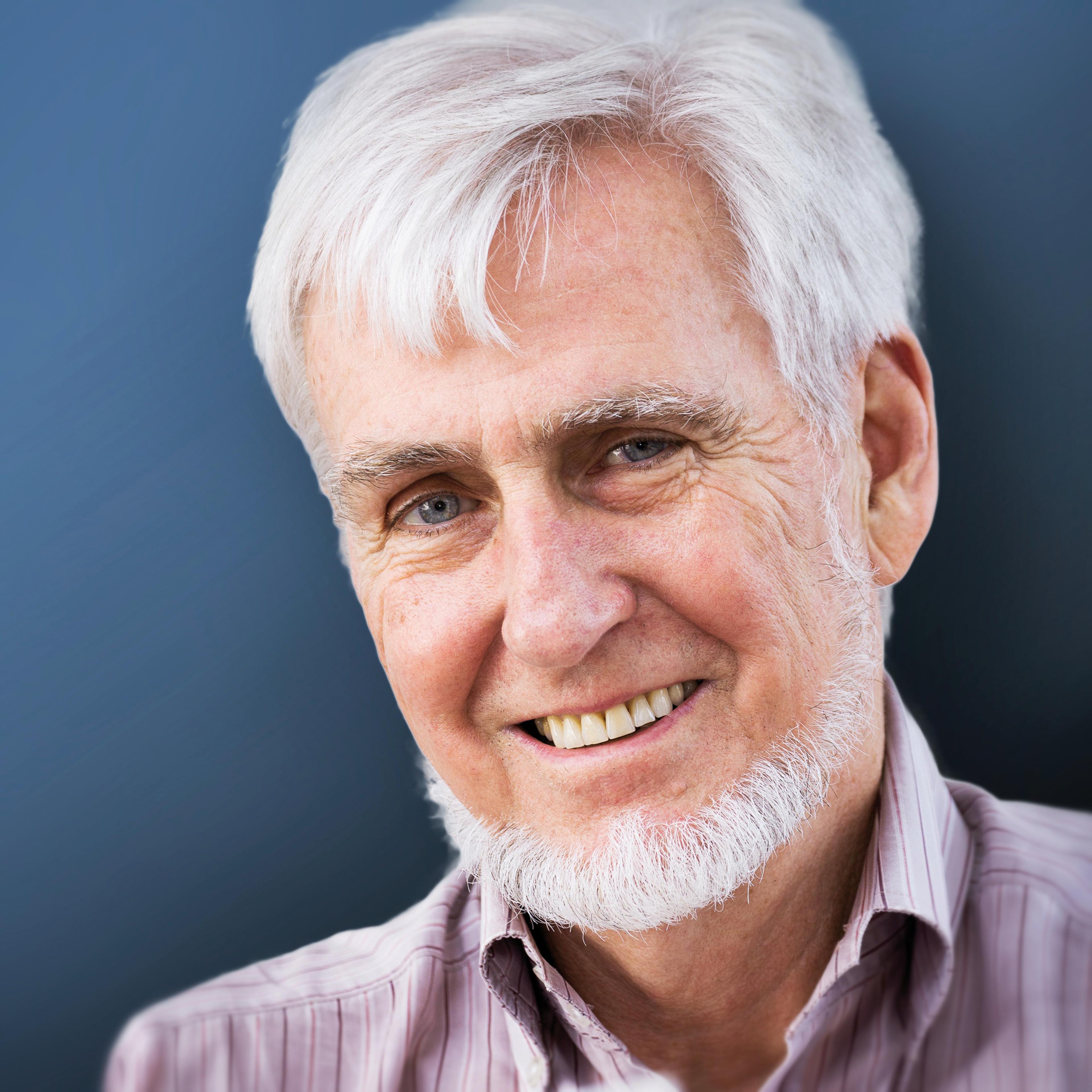
John O'Keefe (Photo credit: © David Bishop, UCL).
His dream to become an aircraft mechanic was thwarted by a shipyard accident during the war. Most of his life was spent as a nightshift mechanic on the New York bus system, where he repaired the buses which became inoperative during the daytime and which couldn’t be fixed by the daytime mechanics. My mother worked as a welder in the Newark shipyards during the war, which reinforced her strong sense of confidence and mastery. Following elementary education at my local Catholic school, I won a scholarship to Regis, the academically ambitious Jesuit high school in Manhattan, while my best friends went to local Bronx schools.
My time at Regis was unsuccessful. I felt like an outsider and never got to grips with Latin and Greek. Four years of poor grades and low test marks left me demoralised and prevented me from getting any help with college fees, which even then were unaffordable. So I went out to work: an entry-level clerical job in a stock brokerage house on Wall Street was followed swiftly by a bookkeeping job in the engineering department of an insurance company. I did not see this as my long-term future and decided to have another crack at the academic world. It was the era of Sputnik, aeronautical engineering was a glamorous profession, and I wanted to put as much distance between myself and the classics as I could.
I spent three years studying aeronautical engineering at New York University in the evening while working in the daytime. After the first year I was lucky to attend a talk by one of the project engineers at Grumman Aircraft Corporation on Long Island, who encouraged me to apply for a job there, which I duly got. I worked on several different aeroplanes, spending considerable time on the shop floor as well as at the drafting board. In the end, however, the gruelling schedule involving over 100 miles commuting a day in rush hour traffic and 12–16 hours of evening lectures per week in addition to a full-time job took its toll and I pined for the freedom of a full-time college student. It was also during this time that I became interested in philosophy through some of my non-engineering courses and decided that many of the perennial problems in philosophy might be solvable through brain research.
City College of New York and McGill University
So in 1960 I give up my job and went back to college full-time. This was a particularly difficult decision since Grumman’s response was to offer me a substantial raise and a role in the development of the Lunar Expedition Module component of the Apollo spacecraft programme, which they were preparing to tender for and eventually were awarded. A path not taken … I was fortunate to be accepted for full-time study at City College of New York, a part of the City University of New York and one of the few tuition-free colleges in the United States. At CCNY, I took courses across a wide range of subjects, paying scant attention to faculty or discipline boundaries. I studied filmmaking, advanced English literature, physics and a wide range of psychology and philosophy courses. I met my wife Eileen in an advanced philosophy course on ethics. There were of course no neuroscience courses in those days, but I was fortunate to take courses in physiological psychology (as it was then called) with Daniel Lehrman, one of the early neuroethologists, and Philip Ziegler, a young enthusiastic researcher just starting his own laboratory.
Phil allowed me to join his team working on the effects of lesions of the wulst on pigeon exploratory behaviour. I got a firsthand taste for experimental brain research and was hooked. During this period I supported myself working in the library, showing classic European films for various courses, and driving a taxi cab in the evening. I loved every minute of it, and had no thought for the future.
Eventually I was summoned to the office of an irate Dean who pointed out that I had accumulated enough credits to receive several degrees and would I please choose one and get out. I opted to major in psychology and minor in philosophy and graduated in 1963. Faced with the unthinkable prospect of working for a living again, I took the advice of one of my professors and applied for graduate school. Again I was lucky and was accepted to study in the McGill University Psychology Department, where Donald Hebb was still active and influential. Hebb was one of the founders of physiological psychology who, in his landmark book The Organisation of Behaviour, provided the theoretical framework which enabled us to think about the neural network basis of cognitive representations.
McGill at that time was the Mecca for the study of physiological psychology. In addition to Hebb, the faculty included Peter Milner, who had discovered rewarding electrical self-stimulation of the brain with Jim Olds, Brenda Milner, whose investigations of the famous patient HM had identified the memory functions of the hippocampus, Wilder Penfield and Herbert Jasper at the Montréal Neurological Institute, who had pioneered electrical stimulation of the brain of conscious patients undergoing surgery for epilepsy, and Ronald Melzack an expert in pain who became my PhD advisor. McGill provided a wonderful environment where students were encouraged to think hard about the brain and creatively about experiments, but where resources were initially extremely limited. Fortunately just after I arrived Melzack received a large grant to study alternative techniques for monitoring brain activity and generously allowed myself and Ken Casey, a postdoctoral fellow who went on to become Head of Neurology at the University of Michigan, to build a state-of-the-art electrophysiological recording laboratory. Ken also taught me the fundamentals of electrophysiology during experiments in which we looked for the midbrain targets of the ascending somatosensory projections. During my final year there I learned a considerable amount of experimental technique from Dr Herman Bouma, who had originally come to work with Hebb on vision but joined me in my amygdala project. On his return to Holland, Herman became the head of the Perceptual Research Laboratory at Phillips, Einthoven where he subsequently had a successful career discovering important principles governing reading.
During my PhD thesis work I developed techniques for recording from chronic animals and concentrated on the amygdala. Jim Olds, although he had left McGill before I got there, was revered there as the co-discoverer of self-stimulation with Peter Milner. In 1966, I learned from Ken Casey, who had visited Olds’ laboratory at Michigan as part of a job interview, that he was successfully recording from single units in awake rats using implanted microwires. I purchased the minimum order of 50,000 feet of coated nichrome microwire, set up a poor man’s version of Olds’ gold-plated lab and improved on his techniques in three ways. Firstly, I used differential recording between adjacent electrodes, which eliminated much of the movement and muscle artefact. Secondly, I introduced the use of preamplifiers made from miniature field effect transistors (which had just become available in 1965) on the animal’s head which greatly improved the signal-to-noise ratio and allowed us to move away from the thick and cumbersome microdot noise reduction cables to lightweight flexible hearing aid wires, greatly improving the animal’s mobility. Finally, I started playing with head-mounted microdrives, first at McGill but then more extensively when I went to London. Many of our early microdrives had four independently movable electrodes and it was only when Caroline Harley came to London on sabbatical and asked for a simpler single drive electrode that I developed the “poor lady’s” single-screw microdrive that we still use extensively and which is now sold by Axona. For the amygdala work, I had recorded from several silent cells for long periods (in some cases days and weeks) before discovering the specific ethological stimulus which caused them to first become active: cells there responded to highly specific stimuli approximating to the classical muchmaligned grandmother cells of Jerry Lettvin. I found mouse detectors, specific food detectors, and bird song detectors. In contrast, the more active cells were sensitive to a broader range of stimuli, for example responding to pure tones of a wide spectrum of audio frequencies. On this basis I formulated my first law of the nervous system, which is that the silent cells are the important ones. I was intellectually and methodologically prepared to tackle the hippocampus and in particular its silent cells. But not quite yet.
Postdoc at University College London
After McGill, Eileen and I decided we wanted to go to Europe and in particular to England. I originally went to University College London in 1967 as a US-NIMH postdoctoral fellow to work on somatosensation with Patrick Wall. We immediately fell in love with Britain. Our first son was born soon after we arrived and, in contrast to our experience in Québec where agencies found us insufficiently religious, the adoption of our second son proceeded smoothly a few years later. British institutions such as the National Health Service, the Ordnance Survey Map with its well-marked walking trails, and the BBC offered a cultural and social landscape that meshed with our lifestyle. University College London also proved the ideal location for me to carry out single unit recording in the freely moving animal. After my NIH Fellowship ran out, I applied for and obtained several grants from the Wellcome Trust and various British research councils which paid my salary as well as providing monies to carry out experiments. This high-risk strategy of funding my own salary as well as my research allowed me to minimise my teaching and administrative duties and maximise research time, and I carried on doing this for my entire career. I got used to letters from the University Human Resources Department advising me to prepare to exit the lab and UCL if my next grant wasn’t funded. Thankfully the emphasis now given to high-impact translational research and rapid, frequent publication had not yet gained ascendancy in those days, allowing for the funding of the risky time-consuming basic research that is my addiction.
I first began recording single units in the hippocampus following an experiment which went astray. The time was 1970 and my project was to record from the dorsal column nuclei during various behaviours in the behaving rat to see whether the descending afferents from the neocortex would modify the excitability of these first-order sensory cells. This was an extremely difficult project, since the juncture of the foramen magnum and the C1 spinal vertebra is designed to be maximally flexible and obtaining decent stable recordings was well-nigh impossible. After two years, the project was clearly not going anywhere. A much easier task would be to record from somatosensory thalamus and neocortex and that’s what I did in my spare time. During one of these experiments I tried to implant a microelectrode in the somatosensory thalamus, but the coordinates I used were too lateral and it strayed into the hippocampus. The first hippocampal cell I recorded was an interneuronal “theta” cell and I was immediately struck by the strong correlation of its activity with the hippocampal sinusoidal 8–10 Hz local field potential theta pattern on one hand and the animal’s motor behaviour on the other. I had previously made LFP recordings from the hippocampus at McGill and was aware of Case Vanderwolf’s claim that theta activity in the rat was correlated with voluntary movements. And here, at the single unit level, was striking confirmation of his claim. I decided then and there to leave the somatosensory system and begin research on the hippocampus. I was intrigued by the apparent conflict between the motor correlates of the cells at single cell level and the widely accepted memory function which had been ascribed to the hippocampus by Brenda Milner on the basis of her research with HM and which I had completely accepted. Brenda had been one of my teachers at the McGill Psychology department and I and many of the other graduate students routinely traipsed across the frozen campus to attend her lectures at the Montréal Neurological Institute.
In addition to the theta unit it was clear that there were other cells in the hippocampus which were mostly silent except for the occasional action potential to announce their presence. Again I was hooked. My amygdala-inspired first law of the nervous system was that silent cells are the important ones and here was a brain region chock-a-block full of them.
I will always be grateful to Pat Wall for allowing me to make this shift to a part of the brain which was outside his immediate field of interest. In addition to his support he signed off on grant applications and provided space for many of the early years of our hippocampal research, turning a deaf ear to the many naysayers who considered the whole approach a waste of time.

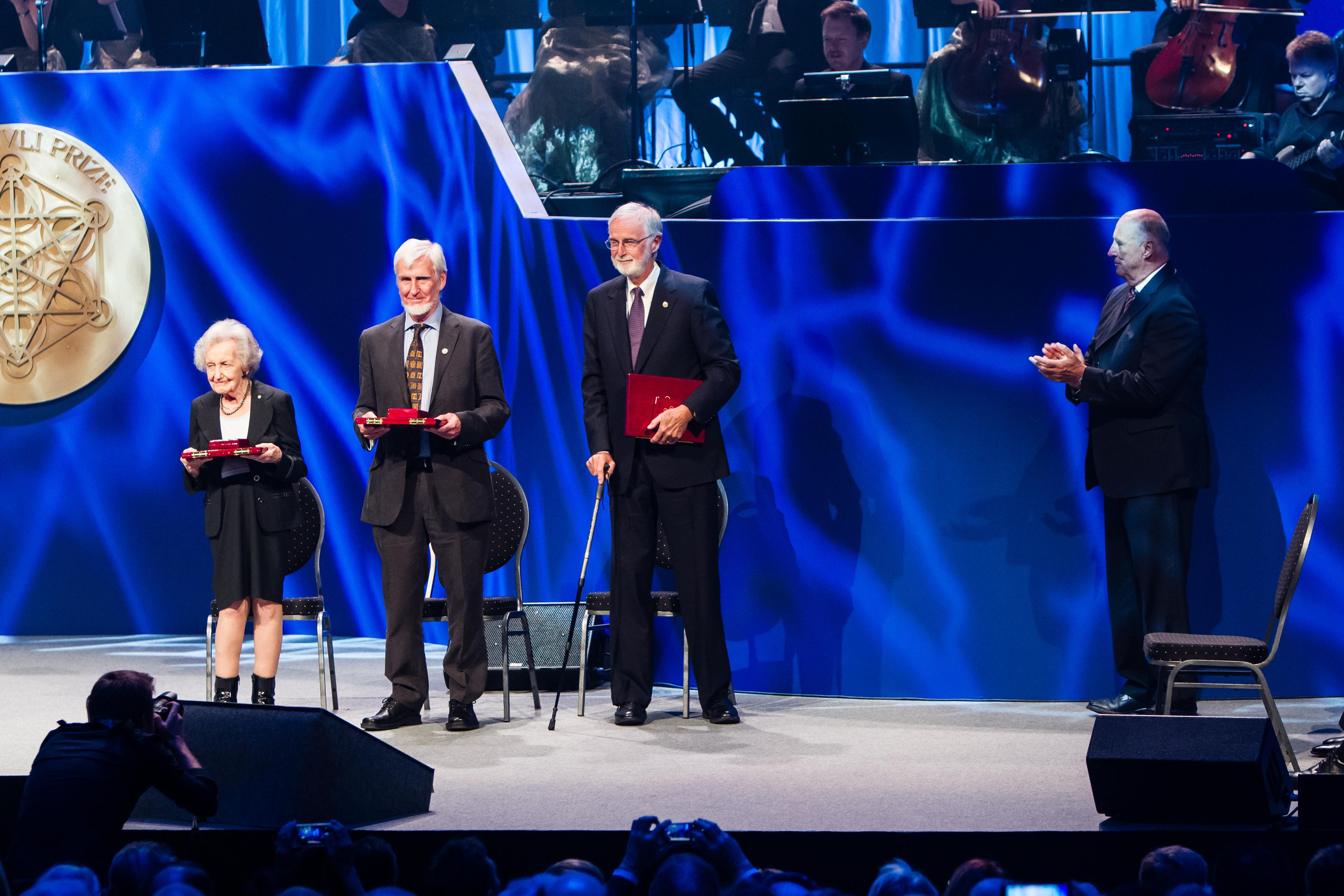
All three 2014 Kavli Prize laureates in neuroscience on stage at the ceremony in Oslo together with His Royal Highness King Harald. From left: Brenda Milner, John O'Keefe and Marcus E. Raichle (Photo credit: Scanpix).
Discovery of the Place Cells
Around this time Jonathan Dostrovsky, an MSc student, joined the laboratory. We recorded single units while the animal was engaged in a wide variety of tasks including basic everyday behaviours such as eating, drinking, grooming, exploring novel environments, searching for foods as well as during simple learned tasks such as lever pressing and approaching different stimuli for food. We noticed two things almost immediately. First, there were two types of cells distinguishable on the basis of strictly physiological properties: spike amplitude and width, and baseline firing rates. As I had originally observed, many of the cells with large amplitude action potentials were silent most of the time, only occasionally showing a burst of spikes when the animal sat quietly or during slowwave sleep. These bursts occurred on large sharp wave spikes in the extracellular hippocampal LFP accompanied by a high frequency waveform which we originally called “wiggles” but quickly changed to the more euphoneous term “ripples.” When we mapped the distribution of these two potentials we found that the ripples were largest in the centre of the CA1 pyramidal cell layer but that the associated sharp wave peaked several hundred microns below in the apical dendrites of the pyramidal cells. More recently, Gyuri Buzsaki, Matt Wilson and Bruce McNaughton have suggested that ripples represent a form of replay of immediately previous spatial learning and might be involved in consolidation of recent memory traces.
It was also immediately obvious that the correlate of the second cell type, which had a much higher resting firing rate and showed a clear phase locking to the ongoing LFP theta oscillations, was a tight coupling to some aspect of movement as I had noticed on my first foray into the hippocampus. This movement correlate was not related to any single limb movement or any specific behaviour but was associated with some higher aspect of the movement such as the vigour or speed with which the movement was executed. Movements which changed the animal’s location seemed to be particularly important. It took much longer for us to identify the correlate of the major cell type, the low firing-rate pyramidal cell. Over a period of months, I began to suspect that their activity didn’t depend so much on what the animal was doing or why it was doing it but had something to do with where it was doing it. And then on one electrifying day I realised with a flash of insight that the cells were responding to the animal’s location or place in the environment. We quickly verified that changing many aspects of the environment one at a time had little effect on the locational response of the cell but if major alterations were made, e.g. by removal of the curtains surrounding the platform, the cell activity altered abruptly. In thinking about these results over the next day I was assailed by a montage of ideas about the potential significance of this finding: the first was that it might mean that the hippocampus was the neural site of Tolman’s cognitive map, a vague hypothetical construct that he had used to explain some aspects of rodent maze behaviour but which had never gained much acceptance in the animal learning field and which was little discussed in the 1960s. It was clear he had given little thought to the neural basis of this ‘map’, much less envisaged that it would be localised in a particular brain structure. This spatial map idea provided a clear function for the movementrelated hippocampal cells and the LFP theta activity, since a map would need information about higher order aspects of an animal’s behaviour such as speed in order to calculate the distance it had travelled. I decided subsequently to christen these movement-related cells “displace cells” to reflect this idea. It also dawned on me that the difficulties that animals with hippocampal damage had in experiments such as those of the Blanchards might be in identifying places in the environment as opposed to objects as the source of threat. The Blanchards had shown that hippocampal-damaged animals could learn to avoid specific threatening objects but were less good at identifying less specific threats such as electric shocks delivered through the floor. Perhaps the hippocampus in rodents was a specialised type of memory system, a memory system for places which, when elaborated, might provide the basis for the more general episodic memory system of the human. Finally, I realised that if the hippocampus were involved in spatial representation we could draw on several millennia of mathematical, philosophical, and geographical thought to help us understand its functions. One of my philosopher heroes had been Immanuel Kant, who had suggested that our sense of space was a special property of the brain which provided a framework for the representation of other aspects of the world such as objects and which existed prior to experience with those objects. Had we found the neural basis for Kant’s a priori spatial faculty of sensibility? Would the hippocampus provide brain researchers with a neural Rosetta Stone, a portal into the mysterious world of cortical brain function? Throughout the day, I experienced a prolonged euphoria of the classical Archimedean type.
I decided to write a short paper on our findings and also to announce the idea that the hippocampus was Tolman’s cognitive map. The paper was originally rejected by Brain Research but after minor modifications finally accepted. I confidently sat back and waited for the chorus of approval from the hippocampal community. Instead there was a deafening silence, with the exception of a small number of isolated voices.
Unbeknownst to me, Jim Ranck at the University of Michigan and subsequently Downstate Medical Center in Brooklyn was carrying out similar recording experiments in the rat hippocampus and finding similar behavioural correlates. His ‘theta’ cells were identical to our displace neurons and his approach-consummate and approach-consummate mismatch cells might be place cells since they consistently fired when the animal approached a reward location on a particular trajectory, i.e., passing through the same place. Ranck did not explore this possibility but on a subsequent sabbatical visit to our laboratory agreed that the animal’s location might be the primary correlate. In subsequent work with Phil Best he confirmed our findings and supported our interpretation. Importantly, he went on to discover the head-direction cells, providing strong support for the cognitive map theory.
The Hippocampus as a Cognitive Map
Around this time Lynn Nadel joined the UCL Anatomy Department to work on the visual system. He quickly became interested in place cells and the cognitive map idea and we decided to write a short review article fleshing out some of the ideas and showing how they applied to the literature on the effects of hippocampal lesions on behaviour. Pat Wall was strongly supportive of the idea, but I’m sure he had no idea how extensive and ambitious the project would become. Our first draft ran to several hundred pages and it was clear that we had a book rather than a review article on our hands. In 1972, we sent the first draft to 50 colleagues and asked for their opinion, and I am still grateful to all those who replied and gave us such constructive comments. However, Oxford University Press, which had agreed to publish the book, also sent the manuscript to reviewers, one of whom was the foremost expert on animal behaviour. He gave it a long, detailed blisteringly negative review, making it clear that we knew little about animal behaviour and that the book suffered badly from this lack of expertise. We could either scrap the project or become experts in animal learning theory and totally rewrite it. We chose to do the latter and found to our amazement that many of the ideas we were expressing about the role of hippocampus in behaviour made a lot of sense within the context of animal learning theory. In total it took us six years to write the book and since many copies of the 1972 version had found their way into the hands and minds of a large number of physiological psychologists, a degree of scepticism developed about whether the book would ever be published at all.
The Hippocampus as a Cognitive Map (HCM) was an ambitious book in conception, daunting to write, and an unavoidably demanding read. The modest OUP run of a few thousand protected many from the effort. It remained cited by many but read by few, until around 2005 when I had it laboriously scanned and made available on the web where it has been and still is free to download.
During the writing of the book, I continued to work on place cells and explore their properties, leading to a more extensive publication in 1976. I reported that in addition to the standard place cell there were other types of spatial cells in the hippocampus including misplace cells which fired maximally when the animal went to a familiar location and found a new object there or failed to find an expected object, and displace cells which Jim Ranck called theta cells because of their close relationship to the ongoing LFP theta and which I characterised as being related to the same aspects of movement as Vanderwolf had attributed to the LFP. In the discussion section of the paper, I speculated that there were two independent ways in which a place cell could be activated. The first was by direct activation from the environmental sensory inputs which impinged upon the animal in a particular location and the second was through a path integration mechanism internal to the hippocampus itself, which used abstract measures of the animal’s behaviour such as its direction and distance of movement since the previous known location to update the representation. This idea has received considerable support since, most recently from work in our lab by Guifen Chen showing that, in a virtual reality environment, 25% of place cells are influenced primarily by the visual inputs while most of the rest receive a significant path integration input as well, i.e. receive a combination of both.
Experimental Test and Support: Lesion Studies
Nadel and I were joined in our analysis of the lesion literature by Abe Black from McMaster University, who came to London every summer and worked with us on ideas about the application of the theory to the normal animal learning literature and the lesion literature. Abe was a world leader in the field of animal learning theory, with a particular interest in avoidance learning, and the three of us published a paper on this in 1975. To our great regret Abe was diagnosed with stomach cancer and died in 1978 at the very young age of 49. I sometimes wonder what the hippocampal field would look like today if he had survived.
What was lacking however was a spatial memory task specifically designed to depend on the capabilities we had attributed to the cognitive map. In the mid-1970s, a newly graduated animal learning theorist, Richard Morris, came to visit Lynn Nadel and myself announcing that he had experienced a Pauline conversion and would like to work on the cognitive map idea. The theory made the strong prediction that hippocampal damage would lead to deficits in allocentric spatial navigation. Lynn and I had tried to develop a land-based navigation task which would be a sensitive test of this hypothesis. The basic idea was that the animal would be started from several locations and had to find a safe location in the environment despite having to move in different directions on each trial to get there. We failed to come up with a workable task, but Richard succeeded. His important idea was to require the animal to go a hidden platform in a swimming pool where there were no local cues to guide it. Together with Nick Rawlins, we subsequently tested rats with hippocampal lesions and found they had pronounced deficits: the Morris water maze was a superb test of hippocampal function and is still the best and most widely used behavioural assay available.
Experimental Test and Support: Single Unit Studies
The Cognitive Map theory also made strong predictions about the existence of other types of spatial information in the hippocampal formation, for example predicting the existence of cells representing distance and direction which would bind together the place representations into a map-like structure. Cells signalling the animal’s heading direction were found by Ranck, Taube, Muller and colleagues in the presubiculum in the 1980s; grid cells in the entorhinal cortex which may be signaling distance travelled in a particular direction have recently been described by the Mosers and their colleagues in 2005. In the early ’80s I was lucky enough to attract Bruce McNaughton and Carol Barnes to spend a year as postdocs in my laboratory. They were already experts in intracellular recording, long-term potential studies and behavioural studies. It was during an earlier visit to Graham Goddard’s (another McGill graduate) lab in Dalhousie that I first met them. Using a minicomputer and an overhead camera head tracking system to record unit activity on an 8-arm maze, we carried out the first quantitative measurement of place fields. We showed for the first time that the firing rate of place cells was dependent on the animal’s speed. Surprisingly, unlike on open platforms where the rat was free to move in all directions, on the behaviourally constraining narrow arms of the radial maze almost all of the cells had unidirectional fields, firing as the animal moved in one direction but not the other. We also implemented an idea of Bruce’s that two electrodes looking at the same cells might enable us to separate action potentials from anatomically close cells by giving us a stereoscopic view. The stereotrode was born to be followed in a short time by the tetrode, a 4 electrode version which is now in wide use.
Over the next few years, our group showed that place cells could learn to distinguish between square and circular enclosures (Colin Lever) and under certain circumstances could do so in an all-or-nothing manner reflective of attractor dynamics (Tom Wills). A particularly revealing experiment during this period was one in which Neil Burgess and I showed that the firing fields of place cells stretched as the enclosure was stretched from a square shape to a rectangle. This led to the idea that one set of inputs to the place cells reflected the distance to one or more walls of the enclosure in particular allocentric directions. As the box was stretched, these inputs maintained their relationship to the opposing walls resulting in expanded fields. The predicted boundary cells were subsequently found in the subiculum by Colin Lever in our lab and in the medial entorhinal cortex by Trygve Solstad in the Moser lab.

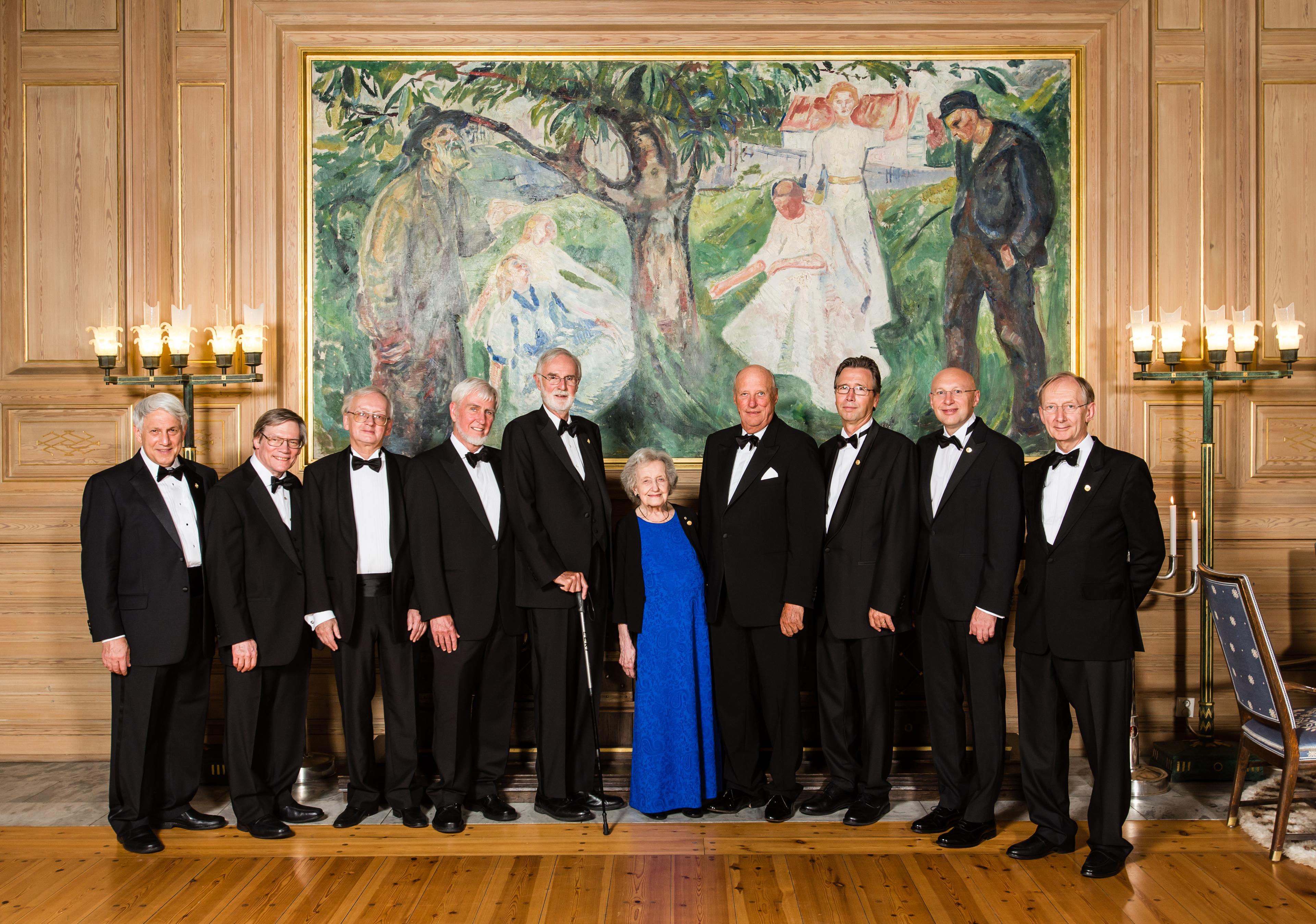
All the 2014 Kavli Prize laureates together with His Royal Highness King Harald in the Munch room at Oslo City Hall at the reception prior to the Kavli Prize banquet. From left: Andrei D. Linde, Alan H. Guth, Alexei A. Starobinsky, John O'Keefe, Marcus E. Raichle, Brenda Milner, His Royal Highness King Harald, Thomas W. Ebbesen, Stephan W. Hell and Sir John B. Pendry. (Photo credit: Thomas Eckhoff).
Phase Precession
For many years, I struggled with the functional role of the hippocampal sinusoidal LFP theta and tried unsuccessfully to integrate it into the cognitive map theory. I knew from my own unpublished work in the late ’70s that at any given time different hippocampal place cells could have different theta phase correlates and even that the same place cell could have different phase correlates at different times. Every so often I would run into Gyuri Buzsaki, who has had a lifetime interest in theta and other hippocampal oscillations and he would ask me what was the relationship of theta to pyramidal cell activity. Given my experience I told him that unlike hippocampal interneurons, they didn’t have a fixed phase of firing relative to theta. I had also been troubled by the possibility that the dentate granule cells might fire like interneurons (it appeared that there were just too many theta cells in the granule layer for them all to be interneurons) and was trying to understand how the interactions between several high-rate dentate granule cells might be translated into low-rate CA3 place cell activity. This got me thinking about interference patterns and led indirectly to my 1985 philosophy paper which suggested that the hippocampus might store and manipulate theta-frequency holograms and that theta was the neural substrate of consciousness (O’Keefe, J. (1985), “Is consciousness the gateway to the hippocampal cognitive map? A speculative essay on the neural basis of mind,” in D.A. Oakley (ed), Brain and Mind, 59–98, Methuen, London.). Around this time I also began exploring the relationship between hippocampal theta sinusoids and vectors. In particular, I pursued the phasor coding idea from engineering in which sinusoids are represented by rotating vectors, but inverted it so that the sinusoids were used to represent vectors and not vice versa. In this version, the length of the vector is represented by the amplitude of the sinusoid and the angle relative to some reference direction is represented by the phase shift relative to a reference sinusoid. This would enable the hippocampus to do vector algebra using sinusoids to represent the locations of environmental landmarks in a polar co-ordinate system (see my paper O’Keefe, J. (1991) “The hippocampal cognitive map and navigational strategies,” in, J. Paillard (ed)., Brain and Space. Oxford University Press, 273–295).
So with all of these ideas in mind, I thought I should have another go at trying to make sense of the relationship between place cells and theta. I went back and looked carefully at the data from one particular place cell from a spatial memory experiment on a +-shaped maze with narrow arms that Andrew Speakman and I had published in 1987. We had also recorded the slow-wave theta LFP from the same electrodes. I quickly confirmed that the phase relationship between the firing of this cell changed from one wave to the next. But how? Was there some systematic relationship between the two? Over several days I looked at run after run on the maze over and over again but couldn’t make any sense of the pattern until one day I found one run on which the spikes fired on every wave and realised that they were systematically moving to earlier phases on each successive wave as the animal ran straight through the field on the narrow track. I also noticed that quite often the phase would continue to precess in the second half of the field despite the fact that the firing rate was falling. This suggested that a simple depolarisation model might not be adequate to explain the effect. Harking back to my thinking about interference patterns, I realised that the wavelet produced by the interference pattern between two waves of slightly different frequencies would produce the required effect. The number of spikes on each theta burst would increase towards the centre of the field and then decrease while the peak of the interference wavelet would continue to progress relative to extracellular theta LFP. I guessed that the phase might correlate with the animal’s location in the field rather than with time or some other variable and this proved to be the case. Following rejection from several journals, the paper by Michael Recce and myself was eventually published in Hippocampus. This was also the first published paper in which tetrodes were used. We considered the possibility that there was a second higher frequency wave in one of the inputs to the hippocampus, so Kate Jeffery looked for it in the entorhinal cortex and Charles King looked for it in the medial septal. Neither found it and we were forced to conclude that if it existed it was located in the dendrites of the pyramidal cells themselves.
Grid Cells
I first met Edvard and May Britt Moser, who later would become my Nobel co-laureates, when they were graduate students in the lab of Per Andersen in Oslo. They were clearly very bright and ambitious and I was more than happy subsequently to agree to their spending some time in my UCL lab to learn the techniques of single unit recording in free-moving animals. Following a string of high-profile important papers on place cells in journals such as Science and Nature, they and their students Torkil Hafting and Marianne Fynn produced their monumental 2005 paper announcing the existence of grid cells in the entorhinal cortex. The grid cells looked like they might provide the metric for the map, completing the spatial information necessary for creating the hippocampal cognitive map. Although others including ourselves had looked in the entorhinal cortex for spatial cells we had all missed the grid cells. I have no doubt that this discovery together with the findings on the spatial role of the human hippocampus have had a major impact on the neuroscientific community’s acceptance of the cognitive map theory. But how were the grid cells constructed? Neil Burgess, Caswell Barry and I suggested that a generalised two-dimensional version of the oscillatory interference model using interference patterns between several theta-like oscillations might provide a good model for the generation of grid cells. And how universal is the grid pattern? Recently Julia Krupic and Marius Bauza in our group have shown that the walls of the environment have a much greater influence on the structure of the grids then had previously been realised, even to the point of destroying the grid pattern in highly structured asymmetrical environments such as trapezoids. We still have a lot to learn about the function of the grids and how these remarkable cells are created by the brain.
Human Hippocampus
One of the major obstacles to the acceptance of the cognitive map theory was the belief that the human hippocampus had a broader function than the spatial one proposed for the rodent hippocampus. The strongest contestant here was the declarative memory theory championed by Larry Squire. Declarative memory theory held that the deficit following hippocampal damage included both factual memories as well as episodic memories for events of the past. The cognitive map extension to the human disagreed with this, predicting that the global memory deficit was limited to episodic memories comprised of memories for what happened in a particular place at a particular time. The idea was that episodes were built upon the basic spatial framework through the addition of a linear sense of time amongst other higher-order cognitive capacities.
One problem in studying the role of the human hippocampus in spatial memory was that most of the tasks had usually been presented on a tabletop or as video displays which, could also be solved by non-hippocampal strategies in particular egocentric ones in which objects were located relative to axes fixed to the eyes, head or trunk as well as by hippocampal-dependent allocentric ones. What was needed was a large-scale environment comparable to the mazes in which rodent spatial navigation was tested. Better still if navigation could be carried out by participants whose heads were immobile, so that their brains could be scanned using the rapidly developing imaging technology. The solution was to use one of the newly developed first-person shoot-em-up virtual reality games which were just becoming available in the ’90s and were all the rage with teenagers. Importantly, some came with editors which allowed the games to be modified and components added or deleted. Neil Burgess, with help from Jim Donnett removed all of the monsters, guns etc. from the game Duke Nukem, which provided an excellent complex 70 x 70 m virtual environment with multiple rooms and pathways, and the layout of which participants could explore and learn to navigate around in a reasonable amount of time. Eleanor Maguire and Neil carried out an imaging experiment in which healthy volunteers had their brains scanned while they navigated between locations using either a cognitive map strategy or a route-finding one in which they followed a series of marked paths, a non-hippocampal strategy. To our delight, the hippocampus and surrounding parahippocampal gyrus become active during mapbased way-finding in virtual reality environments. Importantly there was a good correlation between the accuracy of navigation and the amount of blood flow in the right hippocampus. Subsequently, we used fMRI to study the role of the hippocampus in episodic memory within a virtual reality context and showed that the left hippocampus is more involved than the right. We also studied the spatial and episodic memories of patients with bilateral and unilateral mesial temporal lobe damage in virtual environments and corroborated the imaging work, with the left temporal patients displaying selective recognition deficits in episodic memory and the right showing deficits in spatial memory, object recognition and navigation. The bilateral hippocampal patient showed deficits in both episodic and spatial recognition memory but interestingly not in object recognition memory, suggesting that this deficit in the right temporal-lobectomised patients was due to the additional damage to other areas of the temporal lobe outside the hippocampus. Eleanor Maguire went on to develop these ideas to include the notion that the human hippocampus is involved in the construction of spatial scenes and the prediction of future events. She also showed that the posterior hippocampus of expert and highly practised human navigators (London taxicab drivers) was larger than in the rest of us, including bus drivers who extensively travel the streets of London but along fixed routes.
Another obstacle to the acceptance of the cognitive map theory was the evidence that the left human hippocampus was involved in memory for language and narrative. The problem here was that many psychologists believed that visual-spatial processing was at the opposite end of the cognitive spectrum from language processing, the former held to be represented in a two-dimensional static space while the latter being based on serial processing along a single dimension. How could language be processed and stored in a two- or three-dimensional spatial structure? Nadel and I had suggested that one clue might come from spatial language, which might be easier to store in a spatial structure and, if adequately modelled, might form the basis for much of non-spatial language by metaphorical extension. In follow-up articles, I have explored the use of a specific model of hippocampal spatial function to create a mathematical model underlying the meanings of the spatial prepositions in English, prepositions being one important way in which location and movement are captured. Spatial prepositions are part of the closed class elements of language, are usually limited to around 20 in most languages, and are more or less consistent across languages although not necessarily appearing as independent words in the surface structure. This ‘vector grammar model’ was based on the idea that place fields can be located in an environment on the basis of the distance of the animal’s head from a landmark (usually a wall or boundary of the environment) in a specific allocentric direction (for example the wall of the room between the window and the door) generated by Neil Burgess, Tom Hartley and myself (see above). The vector grammar model postulated that almost all of the prepositions had a primary spatial meaning and these identified the underlying places, objects and vectors connecting them. For example in the phrase “to go from London to New York,” “from” would be represented as the origin or tail of a vector at the place “London” with its head at the place “New York.” The meanings of most prepositions can be described in a similar vector-based fashion.
Translational Possibilities
We are optimistic that our understanding of hippocampus function at the network level will allow us to address the neural basis of neurodegenerative and psychiatric brain diseases. There is evidence that some of the earliest neuropathological manifestations of neurodegenerative diseases such as Alzheimer’s occur in the entorhinal/hippocampal formation. One approach is to create mouse models of some aspects of AD and ask how place cells and other aspects of hippocampal physiology become dysfunctional during disease progression. We already know from work with Francesca Cacucci and Tom Wills that place cells are less able to identify the animal’s current location in these mice and this functional loss correlates with the animals’ inability on spatial memory tasks and its increased amyloid plaque burden. In another related approach, Neil Burgess and Dennis Chan are developing sensitive allocentric spatial tasks as diagnostic tests to look for changes in spatial memory during the early stages of dementia.
The Future
Having spent most of my career as a bench experimenter with a small lab, I have recently accepted the position as Inaugural Director of the Sainsbury Wellcome Centre for Neural Circuits and Behaviour at UCL. As the name implies, the SWC will provide a framework for the study of the neural correlates of perception, emotion, memory and behaviour using the latest techniques in optical and electrophysiological recording to identify the underline neural patterns and optogenetic manipulation of cell activity to control those patterns in a causal manner. It is a thrilling time to be taking on this job, given the numerous possibilities opening up for behavioural and systems neuroscience by these new technologies. So now back to the bench …